Right coronary angiogram in PA cranial view
Right coronary angiogram in PA cranial view
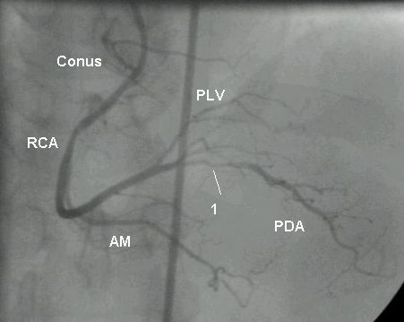
Right coronary angiogram in PA cranial view showing tight lesion in proximal portion of posterior descending artery. PLV: posterior left ventricular branch; AM: acute marginal branch; RCA: right coronary artery. Conus: conus branch of the right coronary artery. Sometimes while cannulating the right coronary artery, the catheter slips into the conus artery. This causes wedging and damping of catheter tip pressure. Prolonged inadvertent injection of contrast into the conus branch can cause ventricular tachycardia or ventricular fibrillation occasionally. A large conus branch crossing the right ventricular outflow tract can cause problem during intra cardiac repair of Tetralogy of Fallot.
Related Posts


About The Author
Johnson Francis
Former Professor of Cardiology, Calicut Govt. Medical Kozhikode, Kerala, India. Editor-in-Chief, BMH Medical Journal