Right coronary lesion and calcification of mitral and aortic valves
Right coronary lesion and calcification of mitral and aortic valves
Coronary angiogram video
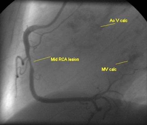
Right coronary angiogram in lateral view demonstrating significant narrowing of mid segment of right coronary artery (RCA). Right ventricular branches are seen just preceding the lesion and distally the RCA divides into posterior left ventricular branches (PLV or PLB) and posterior descending coronary artery (PDA). Dense calcification of the aortic valve is seen to the left of the RCA ostium while mitral calcification is seen in near the distal termination of RCA as PLV.
Such dense calcification indicates that this was a diagnostic coronary angiogram done prior to valve replacement as is usual when the person is above forty years (above 35 years if there are coronary risk factors). In this case it would be a case for double valve replacement with possibly a graft to the right coronary artery.
About The Author
Johnson Francis
Former Professor of Cardiology, Calicut Govt. Medical Kozhikode, Kerala, India. Editor-in-Chief, BMH Medical Journal