Saphenous vein graft (SVG) to diagonal branch of LAD
Saphenous vein graft (SVG) to diagonal branch of LAD
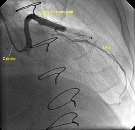
Angiogram demonstrating saphenous vein graft to the diagonal branch of left anterior descending coronary artery in the lateral view. The graft is seen joining the diagonal and the dye flows from the diagonal back into the parent left anterior descending coronary artery. Sternal wires are seen to the left of the image as it is a post coronary artery bypass graft study. Catheter is seen engaging the aortic ostium of the graft directly. Before proceeding with graft angiography, it is always better to look at the operative notes to find the probable site of the grafts. If the notes are not available, sometimes the grafts can be engaged just by probing the aorta with the catheter tip. If this fails, an aortogram can be obtained with a pigtail catheter to locate the sites of origin of the saphenous vein grafts. Once the aortic origin of the graft is located by non-selective aortogram, then it is often easy to directly cannulate the graft ostium to get better graft angiograms.
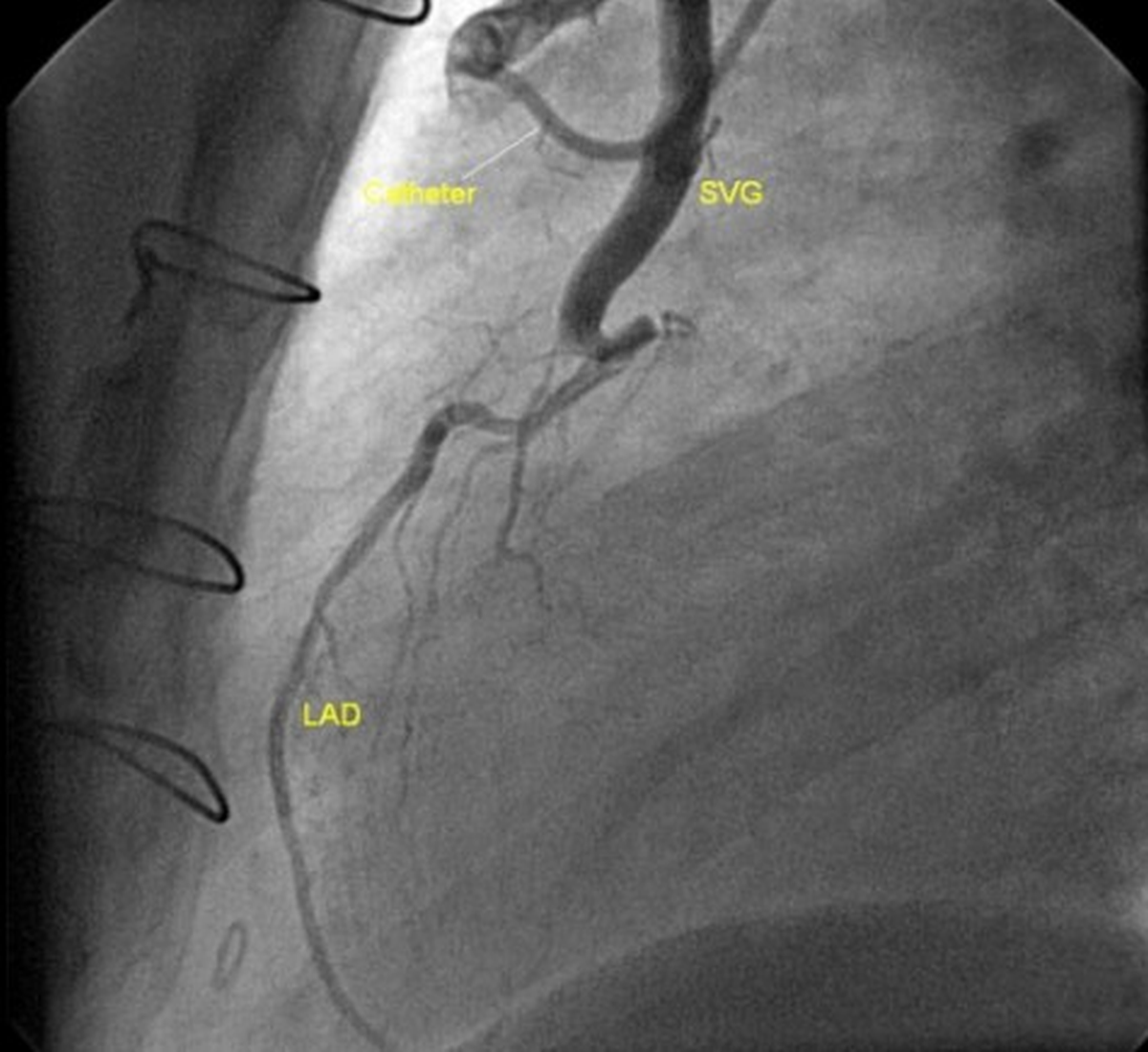
The saphenous vein graft is seen joining the diagonal branch and the dye flows retrogradely into the LAD. The proximal portion of the LAD is seen to be occluded. The sternal wires are prominently seen in the middle of the field as it is a right anterior oblique RAO view. LAD is identified by the septal branches arising from it. It is a type III LAD as it curves around the apex. A type I LAD stops short of the apex and the type II LAD reaches up to the apex. The catheter used is possibly Judkins right coronary catheter.
About The Author
Johnson Francis
Former Professor of Cardiology, Calicut Govt. Medical Kozhikode, Kerala, India. Editor-in-Chief, BMH Medical Journal