Split screen echocardiographic image in parasternal long axis view showing aortic regurgitation jet
Split screen echocardiographic image in parasternal long axis view showing aortic regurgitation jet
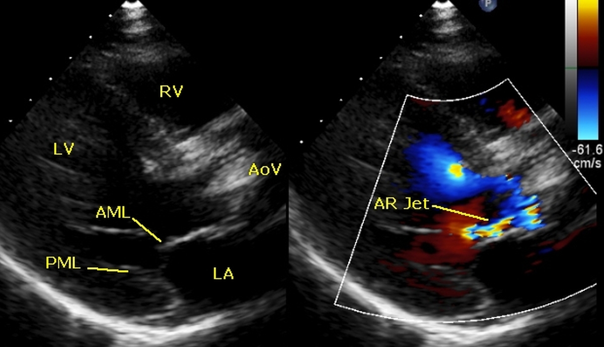
Left panel shows 2-D (two dimensional) image in early diastole indicated by the mitral leaflets (AML: anterior mitral leaflet and PML: posterior mitral leaflet) in partially open position. LV: left ventricle, RV: right ventricle; LA: left atrium. Aortic valve (AoV) is seen as grossly thickened and calcified. The right panel shows the multi coloured (mosaic) jet of aortic regurgitation (AR Jet) along the ventricular surface of the AML. It may be noted that the AML forms the posterior boundary of the left ventricular outflow tract.
Mosaic nature of the colour jet indicates high velocity and turbulence, which is usual with aortic regurgitation as the diastolic pressure in the left ventricle is low while that in the aorta is high, producing a high pressure gradient.
Updated on 1st May 2020
Parasternal long axis view is usually the first view which is obtained in echocardiography. It is obtained by placing the echo transducer in the lower left parasternal region in such a way that the echo beam transects the heart in its long axis from the base to apex. Parasternal short axis view is obtained rotating the transducer ninety degrees. Actual transducer position and angulations are optimized by looking at the image displayed in the screen by the operator. Novices often get views in unusual angulations which make interpretation difficult. Sometimes angulated views are needed when right ventricular inflow needs to be imaged. But usually right ventricular inflow is assessed from the apical four chamber view.
About The Author
Johnson Francis
Former Professor of Cardiology, Calicut Govt. Medical Kozhikode, Kerala, India. Editor-in-Chief, BMH Medical Journal