Parasternal long axis view in normal echocardiogram
Parasternal long axis view in normal echocardiogram
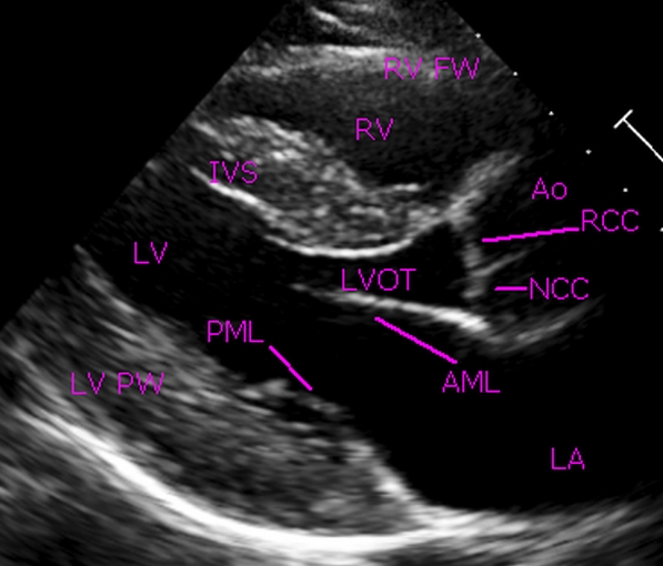
Parasternal long axis view in normal echocardiogram: RV FW: Right ventricular free wall; RV: Right ventricle; IVS: Interventricular septum; Ao: Aorta; RCC: Right coronary cusp of aortic valve; NCC: Non coronary cusp of aortic valve; LVOT: Left ventricular outflow tract; AML: Anterior mitral leaflet; PML: Posterior mitral leaflet; LA: Left atrium; LV: Left ventricle; LV PW: Left ventricular free wall.
Parasternal long axis view is often the first view taken during two dimensional echocardiography. It is taken from the left parasternal region with the ultrasound beam in plan parallel to the base-apex (long axis) of the heart. If the transducer is turned ninety degrees from this view, the parasternal short axis view will be obtained. The most superficial part of the heart imaged in this view is the pericardium covering the right ventricular free wall. M-Mode measurements of the left ventricle, aorta, left atrium and the mitral valve are usually guided from this view. This view is good for visualization of mitral and aortic regurgitations on color Doppler imaging. Doming of mitral and aortic valves in stenotic conditions will be seen well in this view. A perimembranous ventricular septal defect can be seen in this view just below the aortic valve. Aortic override in Fallot’s tetralogy is well seen in parasternal long axis (PLAX) view. Prolapse of the aortic and mitral valve can be seen in PLAX view. Asymmetric septal hypertrophy and systolic anterior motion of the mitral valve in hypertrophic obstructive cardiomyopathy are documented in PLAX view.
Mitral aortic continuity is noted in this normal echocardiogram. There is no intervening tissue between the base of the anterior mitral leaflet and the aortic annulus. Mitral aortic discontinuity is an important feature to look for in certain congenital heart diseases. Wide separation between the two mitral leaflets is well seen in this diastolic frame.
Read more on: Normal adult echocardiography – parasternal views
Related Posts

About The Author
Johnson Francis
Former Professor of Cardiology, Calicut Govt. Medical Kozhikode, Kerala, India. Editor-in-Chief, BMH Medical Journal