Takayasu arteritis
Takayasu arteritis
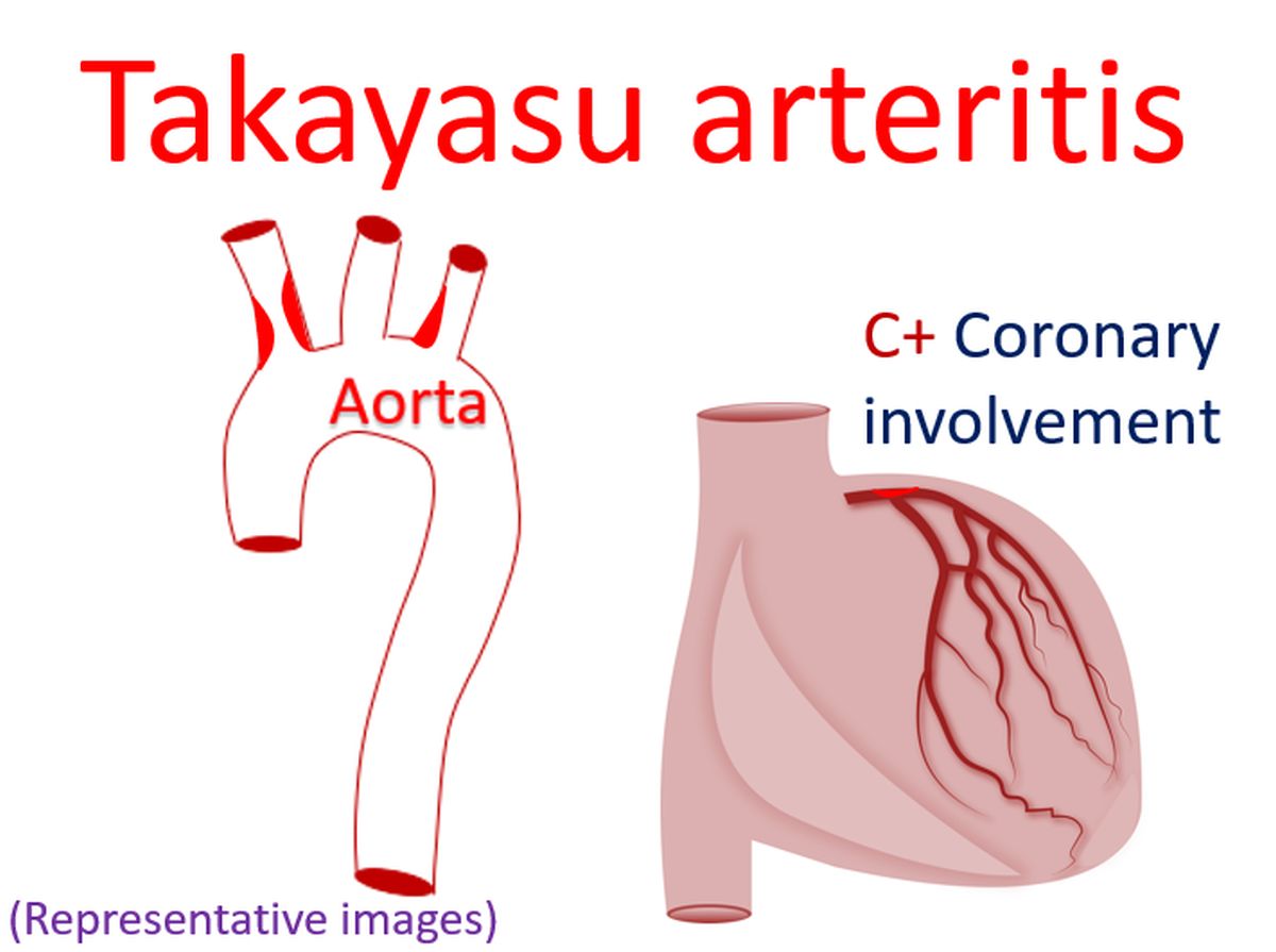
Takayasu arteritis is an inflammatory disorder affecting aorta and its major branches. It is also known as aortoarteritis and pulseless disease [1]. Arteritis leads to thickening of vessel wall, fibrosis, stenosis and thrombus formation. Severe inflammation may weaken the arterial media and lead to aneurysm formation [2].
Takayasu was an ophthalmologist, who noted characteristic fundal arteriovenous anastomoses in a young female in 1905 and published it in Acta of the Opthalmic Society of Japan in 1908 (12:554–5). Onishi and Kagosha described similar cases associated with absent radial pulses in the same year [1].
A study by Lupi-Herrera E et al reported 107 cases over a 19 year period [3]. In their series, females were predominantly involved (8.5:1). Acute inflammatory phase with systemic and cardiovascular symptoms were noted in half of the cases. 65% had supra aortic trunks and abdominal aortic involvement (Type III in the classification prevalent then). Reduction in amplitude of peripheral pulses was noted in 96%, vascular bruit in 94%, hypertension in 72% and renal artery stenosis in 62%. Heart failure was noted in 28%, but rarely due to coronary arteritis. 48% had previous or current active tuberculosis.
Another natural history study published in 1989 had 88 patients [4]. The cumulative survival of Takayasu arteritis at 5 years was 91% of which event free survival was 74.9%. Ten year survival of Takayasu’s arteritis was 84% of which 64% was event free survival.
Ishikawa clinical classification (1978) of Takayasu arteritis had three groups, with two subgroups for group II [5]. Group I had uncomplicated disease, with or without pulmonary artery involvement. Group IIa had mild/moderate single complications. Group IIb had severe single complication. Group III had two or more complications.
An angiographic classification was published in 1997, which had types I to V with two subtypes for type II [6]. This was formulated after a retrospective review of 102 Indian and 80 Japanese patients with Takayasu arteritis, comparing their clinical manifestations and angiographic findings.
References
- Johnston SL, Lock RJ, Gompels MM. Takayasu arteritis: a review. J Clin Pathol. 2002 Jul;55(7):481-6.
- Numano F, Okawara M, Inomata H, Kobayashi Y. Takayasu’s arteritis. Lancet. 2000 Sep 16;356(9234):1023-5.
- Lupi-Herrera E, Sánchez-Torres G, Marcushamer J, Mispireta J, Horwitz S, Vela JE. Takayasu’s arteritis. Clinical study of 107 cases. Am Heart J. 1977 Jan;93(1):94-103.
- R Subramanyan, J Joy, K G Balakrishnan. Natural History of Aortoarteritis (Takayasu’s Disease). Circulation. 1989 Sep;80(3):429-37.
- Ishikawa K. Natural history and classification of occlusive thromboaortopathy (Takayasu’s disease). Circulation. 1978 Jan;57(1):27-35.
-
Moriwaki R, Noda M, Yajima M, Sharma BK, Numano F. Clinical manifestations of Takayasu arteritis in India and Japan–new classification of angiographic findings. Angiology. 1997 May;48(5):369-79.
About The Author
Johnson Francis
Former Professor of Cardiology, Calicut Govt. Medical Kozhikode, Kerala, India. Editor-in-Chief, BMH Medical Journal