Traffic jam sign in constrictive pericarditis
Traffic jam sign in constrictive pericarditis
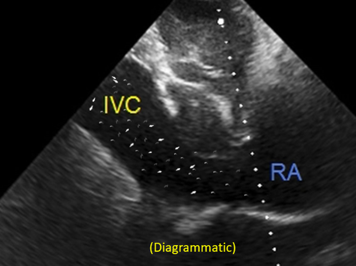
Multiple echogenic shadows are seen moving to and fro in the hepatic veins and inferior vena cava in cases of constrictive pericarditis and is called the traffic jam sign. Same pattern, in a lesser degree, may be seen rarely in other cases of grossly elevated right atrial pressures with dilated inferior vena cava and hepatic veins.
The sign has also been described in a case of critical valvular pulmonary stenosis with delayed presentation by Ramachandra Barik, Siva Prasad Akula and, Sheshagiri Rao Damera [1]. It was a 39 year old man who presented with severe valvar pulmonary stenosis with right heart failure as evidenced by enlarged liver, elevated jugular venous pressure, anasarca and cyanosis. His inferior vena cava was dilated (3 cm) and plethoric with less than 25% respiratory variation and marked by traffic jam sign. His hemodynamic status improved remarkably after balloon pulmonary valvotomy which was preceded by dobutamine infusion and other supportive measures. He returned to his routine life three months after the procedure.
Reference
- Ramachandra Barik, Siva Prasad Akula, Sheshagiri Rao Damera. Use of Dobutamine Stress Echocardiography for Periprocedural Evaluation of a Case of Critical Valvular Pulmonary Stenosis With Delayed Presentation. J Cardiovasc Echogr. Apr-Jun 2016;26(2):56-60.
Related Posts


About The Author
Johnson Francis
Former Professor of Cardiology, Calicut Govt. Medical Kozhikode, Kerala, India. Editor-in-Chief, BMH Medical Journal