Triangle of Koch
Triangle of Koch
Triangle of Koch is situated on the right atrial aspect of the inter-atrial septum. It is bounded by the septal tricuspid leaflet, tendon of Todaro and the coronary sinus. The atrioventricular node is located at the apex of this triangle [1]. The initial description of the triangle was by Walter Koch in 1909 [2]. Knowledge of the Koch’s triangle is very important to safely perform radiofrequency catheter ablation within the right atrium. Ablation within the triangle of Koch has a risk of injury to the AV node and complete AV block [3].
Tendon of Todaro is a collagenous subendocardial band in the right atrium. It constitutes a part of the fibrous skeleton of the heart. Tendon of Todaro runs from the central fibrous body to the Eustachian ridge. Macroscopically, tendon of Todaro is visible only in the fetal and infant heart as a white structure. It involutes later and is visible only microscopically in the adult heart. One study found tendon of Todaro visible macroscopically in only 10.8% of the hearts, that too only in the younger specimens [3]. So, in that study, they used a line joining the apex of the Koch’s triangle to the point where the basal edge of the triangle touches the Eustachian ridge. Apex of the triangle was found by transillumination of the membranous septum from the left ventricular side. Eustachian ridge separates the orifice of the inferior vena cava from the coronary sinus [4].
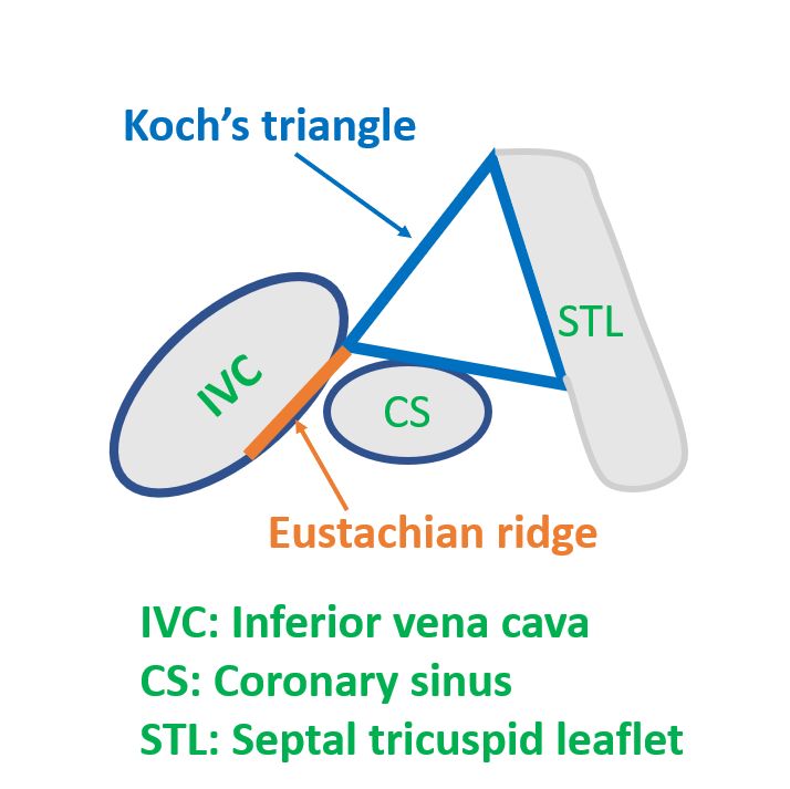
Highly diagrammatic representation of the landmarks used to identify the triangle of Koch is shown above. The boundary of the triangle from the apex to the Eustachian ridge marks the location of tendon of Todaro, seen histologically in the adult and macroscopically in the fetus and infant. It is the region pointed by the blue arrow for marking the triangle of Koch.
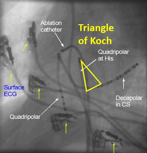
Approximate position of triangle of Koch in the left anterior oblique (LAO) fluoroscopic view is given above. The tip of the quadripolar His bundle catheter marks the approximate position of the apex of the triangle. Proximal portion of the His bundle catheter will be roughly along the Eustachian ridge and tendon of Todaro. Decapolar catheter in the coronary sinus (CS) identifies the base of the triangle of Koch. Tip of the ablation catheter is seen well away from the tip of the His bundle catheter, to avoid ablation in the region of the compact AV node while ablating for supraventricular tachycardia. In case you need ablation of the AV node in case of atrial fibrillation with drug refractory fast ventricular rate, tip of the His bundle catheter will guide the AV nodal location.
References
- Shanubhogue S, Mohamed T, Shankar N. Morphometry of the triangle of Koch and position of the coronary sinus opening in cadaveric fetal hearts. Indian Heart J. 2017 Jan-Feb;69(1):125-128.
- Koch W. Weiter Mitteilungen über den Sinusknoten des Herzens. Verh Dt Ges Pathol 1909;13:85-92; cited in [3].
- Klimek-Piotrowska W, Holda MK, Koziej M, Salapa K, Piatek K, Holda J. Geometry of Koch’s triangle. Europace. 2017 Mar 1;19(3):452-457.
- McKay T, Thomas L. Prominent crista terminalis and Eustachian ridge in the right atrium: Two dimensional (2D) and three dimensional (3D) imaging. Eur J Echocardiogr. 2007 Aug;8(4):288-91.
Related Posts


About The Author
Johnson Francis
Former Professor of Cardiology, Calicut Govt. Medical Kozhikode, Kerala, India. Editor-in-Chief, BMH Medical Journal