Ventricular ectopics – couplets
Ventricular ectopics – couplets
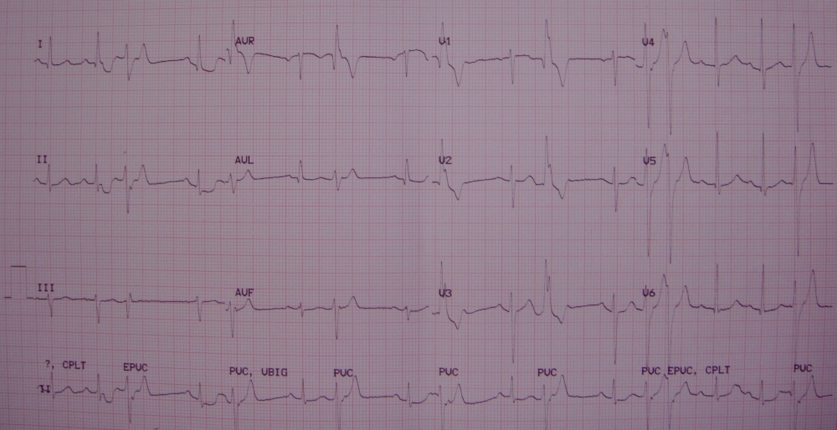
Ventricular ectopic beats in couplets. Couplets indicate a higher potential for arrhythmia than isolated ventricular ectopic beats. If three ventricular ectopic beats occur in a sequence at a rate above 100 per minute is called a salvo or a short run of nonsustained ventricular tachycardia. Upright QRS in V1 [resembling a right bundle branch block (RBBB) pattern] and a slurred S wave in V6 are suggestive of left ventricular origin of the ventricular ectopic beat. Ventricular ectopics are identified by wide bizarre QRS complexes and ST / T changes (ST segment and T wave opposite in polarity to the dominant QRS deflection). They are not usually preceded by a P wave. Sometimes a ventricular ectopic beat occurs just after a usual sinus P wave. Then they are called late diastolic ventricular ectopics and they come in the differential diagnosis of intermittent pre-excitation (WPW syndrome). Alternate possibility which has to be thought of when there are intermittent premature beats with RBBB pattern is supraventricular ectopy with aberrant conduction. In this ECG couplets have been captured in leads V4 – V6 and in the lead II rhythm strip. Other leads have captured isolated ventricular ectopic beats (VPB or VPC). Mild ST segment depression is seen in certain sinus beats in lead I and II. Commonly benign ventricular ectopics originate from the right ventricular outflow tract, while left ventricular ectopy is often associated with structural heart disease.
Related Posts


About The Author
Johnson Francis
Former Professor of Cardiology, Calicut Govt. Medical Kozhikode, Kerala, India. Editor-in-Chief, BMH Medical Journal