Ventricular Septal Defect (VSD) – hole in the wall between the lower chambers of the heart
Ventricular Septal Defect (VSD) – hole in the wall between the lower chambers of the heart
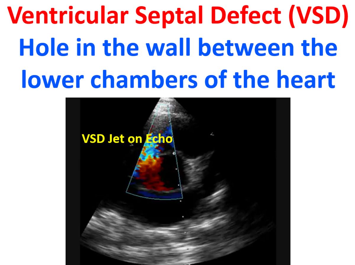
Ventricular Septal Defect (VSD) is a defect in the interventricular septum separating the two ventricles of heart (lower chambers). It is the commonest congenital heart defect. The commonest variety of VSD is the perimembranous VSD situated near the membranous part of the interventricular septum.

Defects can also occur in the muscular portion of the septum. Other sites are inlet VSD (near the inflow portion of the ventricles, near the mitral and tricuspid valves) and outlet VSD near the outflow region below the semilunar valves. VSDs are also divided into supra cristal and infracristal, in relation to the crista supraventricularis, a muscular ridge in the outflow region of right ventricle.
Ventricular septal defect causes a high velocity jet of blood to flow from left ventricle to right ventricle as the pressure in the left ventricle is higher. If the ventricular septal defect is large the left to right shunt is high and can cause overloading and failure of the left ventricle. Large ventricular septal defect is an important cause of heart failure in infancy. Large ventricular septal defects can cause rapid rise of pressure in the pulmonary circulation. Hence these defects have to be closed in infancy itself.

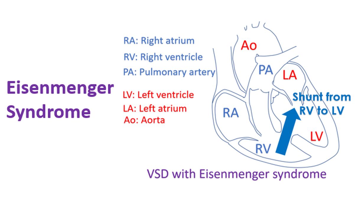
Otherwise irreversible changes in the pulmonary vessels can lead to severe pulmonary hypertension and reversal of shunt from right ventricle to left ventricle. This situation is known as Eisenmenger syndrome and is associated with deoxygenated blood reaching the general (systemic) circulation. Cyanosis (bluish discolouration of skin and lips) occur in such a situation.
Ventricular septal defects can decrease in size over a period and become small or disappear. Small defects do not generally require surgical closure. Large defects, especially those which cause pulmonary hypertension have to be closed early. Surgical closure of VSD is an open heart procedure.
Several of the VSD’s, especially the muscular VSDs can be closed by interventional procedures using devices. A guide wire is introduced through a small skin puncture in the groin and threaded up the femoral artery into the aorta and left ventricle. The wire then crosses the VSD into the right ventricle. The wire is snared out of the body through a catheter (small tube) introduced through the jugular vein in the neck. The sheath for delivering the VSD closure device is threaded over the wire across the defect.

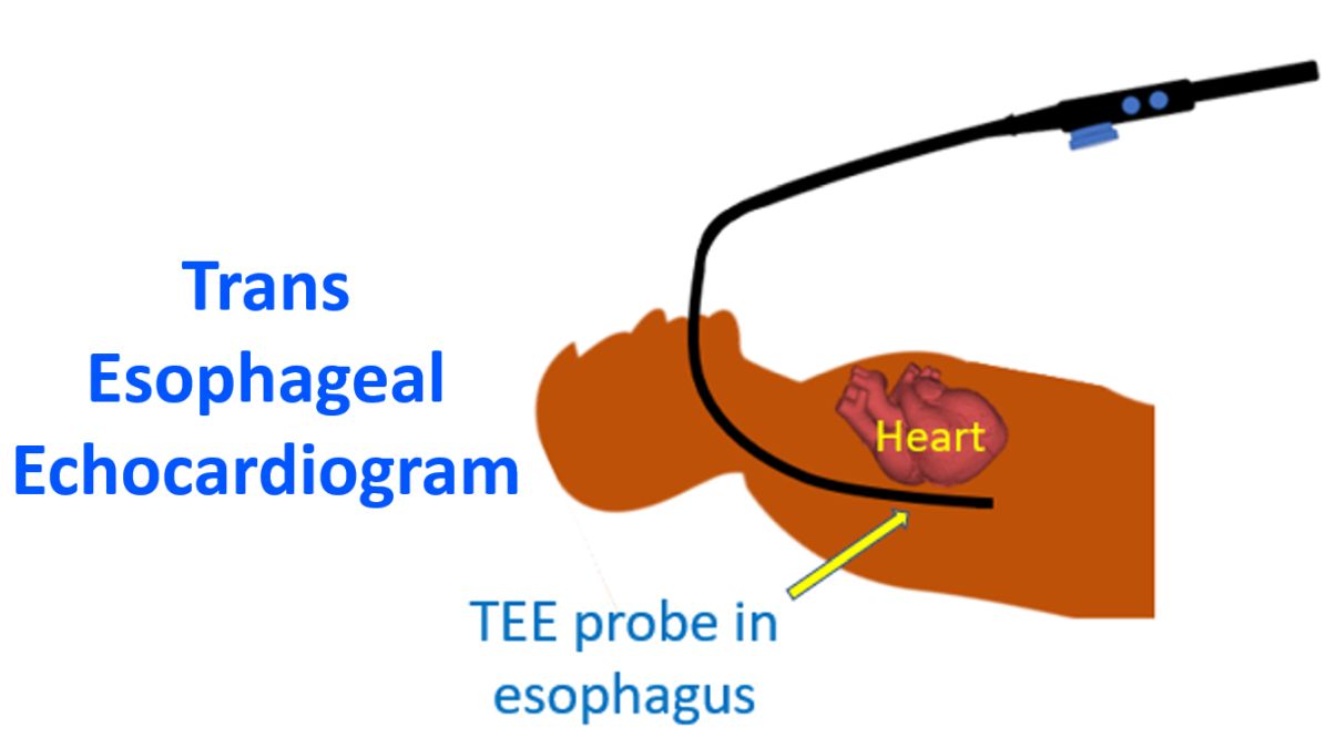
The VSD closure device is threaded through the sheath and delivered across the defect, confirming its position by X-ray fluoroscopy and transesophageal echocardiography (ultrasound imaging using a probe introduced through the food pipe). The person who has undergone VSD device closure can walk about the very next day and go home the day after. There will be only two tiny skin puncture wounds which will seal off automatically. The procedure can be done under local anaesthesia in an adult while children require general anaesthesia.