Accelerated Idioventricular Rhythm
Accelerated Idioventricular Rhythm
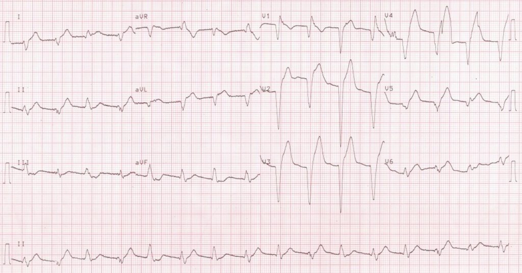 Accelerated Idioventricular Rhythm (AIVR) is a ventricular rhythm originating from the Bundle of His, the Purkinje system or the ventricular myocytes. It can be diagnosed when three or more ventricular ectopic beats occur consecutively at a rate more than the usual idioventricular rhythm which has a rate of 20 – 40 beats per minute. The rate of AIVR is usually similar to that of the underlying sinus rhythm which it supersedes. There can be features of AV dissociation like fusion beats or capture beats. AIVR is often well tolerated and seldom needs any specific treatment. It can be overridden by accelerating the sinus rhythm with atropine. In the thrombolytic era, AIVR was often noted as a reperfusion arrhythmia. An article on the historical aspects of AIVR has been published in IPEJ [1]. AIVR can also occur after spontaneous recanalization of the coronary artery as well as after successful primary angioplasty.
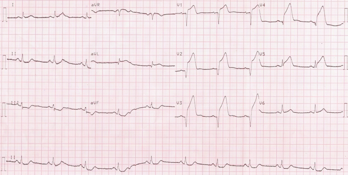 Baseline ECG in this case showing hyperacute ST elevation anterior wall myocardial infarction with Q waves and ST segment elevation in anterior leads (Pardee’s sign [2]) and reciprocal ST segment depression in inferior leads.
AIVR has been associated with drugs like halothane, desflurane, cocaine and digitalis [2].
Idiopathic AIVR
Idiopathic AIVR originating from the right bundle branch has been described as an unusual type of ventricular arrhythmia [3]. Two of their 8 patients had syncope and 3 had left ventricular dysfunction. Metoprolol was useful in decelerating the arrhythmia and relieving symptoms. Five patients underwent electrophysiological study and catheter ablation. Right bundle branch block morphology was noted in sinus rhythm after ablation. Left ventricular function normalized after ablation and symptoms were controlled.
References
- Andres Ricardo Perez Riera, Raimundo Barbosa Barros, Francisco Daniel de Sousa, Adrian Baranchuk. Accelerated idioventricular rhythm: history and chronology of the main discoveries. Indian Pacing Electrophysiol. J. 2010;10(1):40-48.
- Pardee HEB. An electrocardiographic sign of coronary artery obstruction. Arch Intern Med 1920; 26: 244– 257.
- Minglong Chen, Kai Gu, Bing Yang, Hongwu Chen, Weizhu Ju, Fengxiang Zhang, Gang Yang, Mingfang Li, Xinzheng Lu, Kejiang Cao, Feifan Ouyang. Idiopathic accelerated idioventricular rhythm or ventricular tachycardia originating from the right bundle branch: unusual type of ventricular arrhythmia. Circ Arrhythm Electrophysiol. 2014 Dec;7(6):1159-67.
Related Posts

About The Author
Johnson Francis
Former Professor of Cardiology, Calicut Govt. Medical Kozhikode, Kerala, India. Editor-in-Chief, BMH Medical Journal