Apical five chamber view in echocardiography
Apical five chamber view in echocardiography
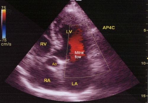
Apical five chamber view is useful in assessing all four chambers along with the aortic valve. LV: left ventricle; Ao: aorta; RV: right ventricle; LA: left atrium; RA: right atrium. The forward mitral flow in diastole is encoded in red colour as it is towards the transducer kept at the apex of the heart. The colour bar on the left upper corner shows the colour coding scheme with blue away from the transducer and red towards the transducer. The colour variance is also shown. The maximum velocity which can be imaged with the particular setting is known as the Nyquist limit, which in this case is 75 cm/sec. Nyquist limit will depend on half the pulse repetition frequency. The graduations on the right side indicate the depth of the image which is about 15 cm from the surface in this case. Mitral and aortic regurgitation as well as stenotic jets can be imaged well in this view.
Related Posts

About The Author
Johnson Francis
Former Professor of Cardiology, Calicut Govt. Medical Kozhikode, Kerala, India. Editor-in-Chief, BMH Medical Journal