Mitral valve prolapse with regurgitation – echocardiogram
Mitral valve prolapse with regurgitation – echocardiogram
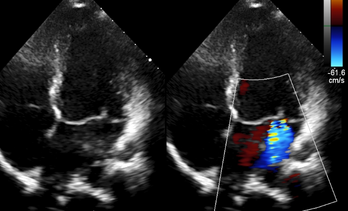
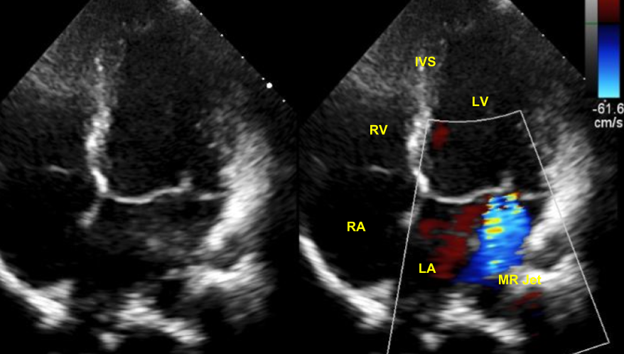
Echocardiogram in apical four chamber view demonstrates prolapse of both anterior and posterior mitral leaflets. The central part of the leaflets can be seen more towards the left atrium than the coaptation point. Mosaic colour Doppler jet of mitral regurgitation can be seen in the left atrium, starting from the mitral valve.
Severity of mitral regurgitation can be estimated by comparing the mitral regurgitation jet area with that of the left atrium. The higher the ratio of MR Jet to left atrium, the higher the severity of mitral regurgitation. Another method is by measuring the width of vena contracta [1]. Vena contracta can be measured in multiple planes. Biplane vena contracta gives a better correlation with regurgitant volumes. Biplane vena contracta width of 0.5 cm or more indicates a regurgitant volume of more than 60 ml and corresponds to regurgitant orifice of more than 0.4 sq cm.
Regurgitant volumes can be calculated using pulse Doppler at mitral annulus and left ventricular outflow tract. Velocity time integral at each site multiplied by the area by planimetry gives the flow. Difference in the flow volumes between mitral and left ventricular outflow tract is a measure of the regurgitant volume. Regurgitant volume divided by the velocity time integral of the MR jet will give the effective regurgitant orifice.
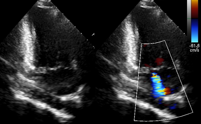
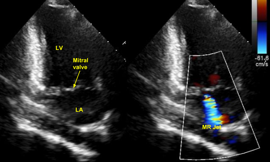
MVP-MR Apical two chamber view
Echocardiogram in apical two chamber view demonstrates the prolapse of both anterior and posterior leaflets as well the colour jet of mitral regurgitation. Scallops of the mitral leaflets are also visible on close scrutiny. Identifying the scallops involved is useful during surgical repair of the mitral valve. This is better achieved by transesophageal echocardiography, especially with real time three dimensional (4D) imaging. 3-D imaging can give a “surgeon’s view” of the mitral valve from both the ventricular and atrial aspects.
Anterior leaflet has three divisions named A1, A2 and A3. Posterior leaflet has three divisions named P1, P2 and P3. Since P1 scallop is near the anterolateral commissure, it is known as the anterolateral scallop. P3 is near the posteromedial commissure and is known as posteromedial scallop. P2 is known as the central scallop [2].
References
- Hall SA, Brickner ME, Willett DL, Irani WN, Afridi I, Grayburn PA. Assessment of mitral regurgitation severity by Doppler color flow mapping of the vena contracta. Circulation. 1997 Feb 4;95(3):636-42.
- Irvine T, Li XK, Sahn DJ, Kenny A. Assessment of mitral regurgitation. Heart. 2002 Nov;88 Suppl 4:iv11-9.
Related Posts


About The Author
Johnson Francis
Former Professor of Cardiology, Calicut Govt. Medical Kozhikode, Kerala, India. Editor-in-Chief, BMH Medical Journal