ECG findings in atrial pacing
ECG findings in atrial pacing
Each P wave is preceded by a sharp pacing spike, indicating atrial pacing with regular pacing and capture. The P wave morphology in atrial pacing is different from the usual sinus rhythm P wave as the sequence of atrial activation is different for atrial pacing compared to sinus rhythm. If the pacing is from the upper part of the right atrium, P waves will be upright in inferior leads as the activation proceeds downwards towards the inferior leads. But if the pacing is from low atrium, P waves will be inverted in inferior leads as the activation proceeds from below upwards in the atrium.
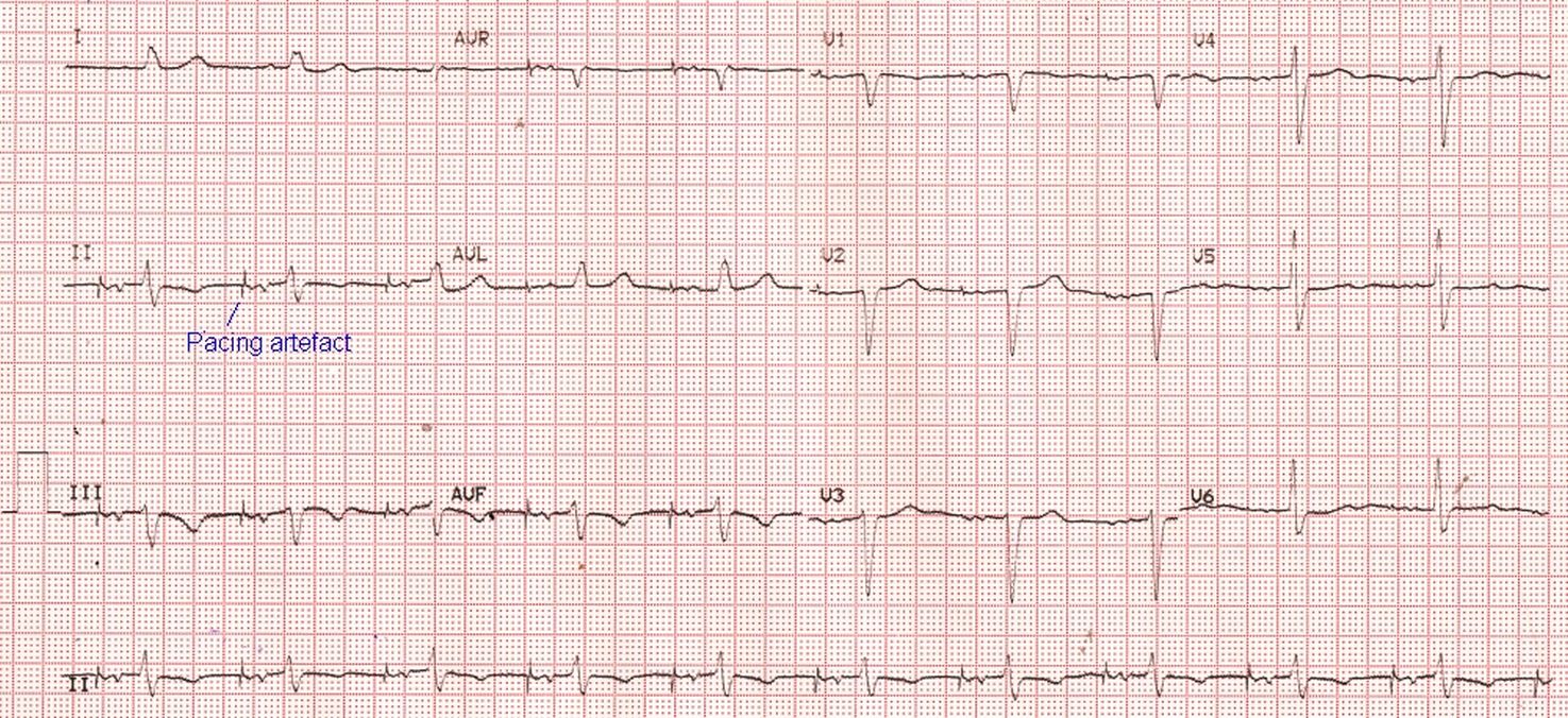
Latency or the interval between the pacing spike and the P wave, can be increased due to atrial conduction disease. It will also widen the P wave. Short and sharp P waves are noted in atrial septal pacing. In upper septal pacing, P waves are positive in inferior leads and V1 will show a small negative deflection. Lower septal pacing on the other hand shows negative P waves in inferior leads and a positive P wave in V1 [1].
Reference
- Das A. Electrocardiographic features: Various atrial site pacing. Indian Heart J. 2017 Sep-Oct;69(5):675-680. doi: 10.1016/j.ihj.2017.08.030. Epub 2017 Sep 1. PMID: 29054201; PMCID: PMC5650557.
Related Posts


About The Author
Johnson Francis
Former Professor of Cardiology, Calicut Govt. Medical Kozhikode, Kerala, India. Editor-in-Chief, BMH Medical Journal