Basic echocardiographic views
Basic echocardiographic views
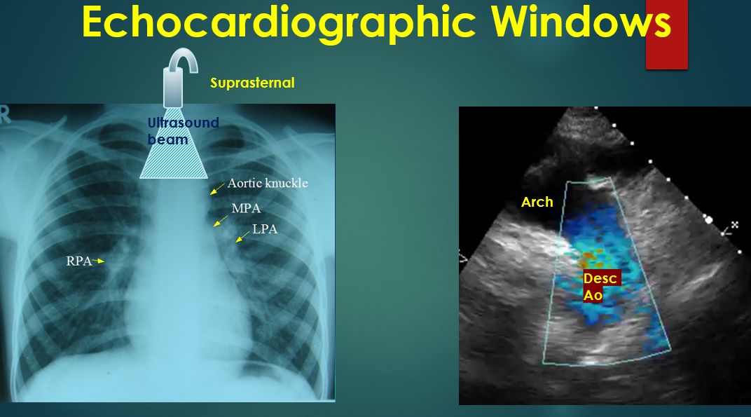
Echocardiography is now not restricted to the echocardiographic laboratory. It is used in the emergency department, at bedside, in the intensive care unit as well as in the operating room. Hence a basic knowledge is needed for all physicians and paramedics. Transthoracic echocardiography is often done from four echo windows. Echo windows are regions on the chest which permit imaging of the heart with least covering by the lungs.
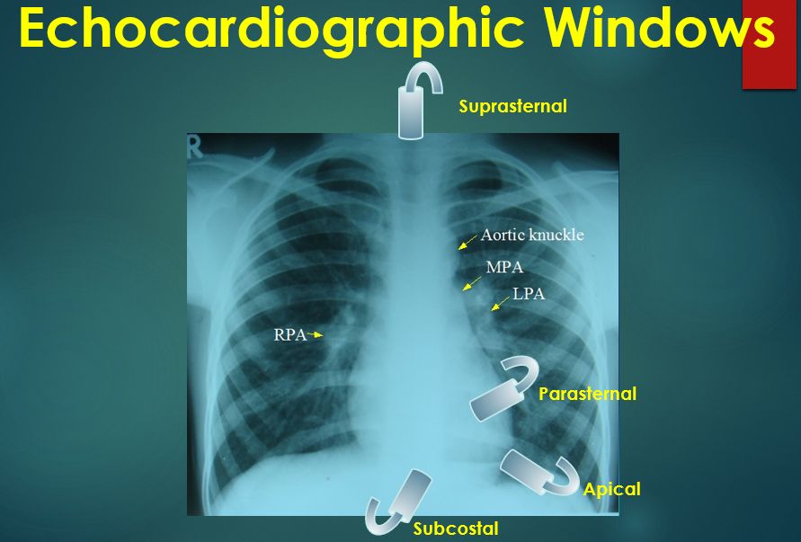
The four common locations at which the echocardiographic transducer is placed for imaging are the parasternal, apical, subcostal, and suprasternal. Parasternal views are often obtained first, followed by apical, subcostal, and suprasternal. A good knowledge of the anatomy of the heart is needed for interpretation of images from each view. This becomes more difficult in complex congenital heart diseases where the cardiac chamber positions and size may vary.
First view to be obtained is often the parasternal long axis view. This view images the heart from the base to apex long axis view. Transducer is placed in the left parasternal region and fine adjustments in angulation are made till a view similar to that in the left panel is obtained. Exact position and angulation will vary between individuals.
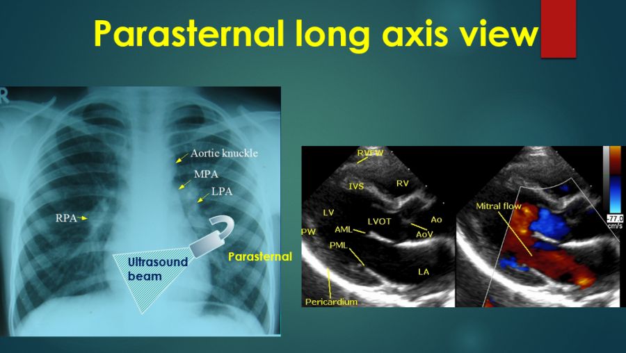
Usual structures imaged in this view are the right ventricular free wall and outflow region, interventricular septum, aorta, and aortic valve, left ventricular outflow tract, anterior and posterior mitral leaflets, left ventricular cavity, posterior wall of left ventricle and left atrium. Colour Doppler imaging is used to image the flow directions and abnormal flows if any.
Here is an animated view from the parasternal long axis. Opening and closing movements of the aortic and mitral valves are visible. Contraction of each region of the left ventricle is also inspected closely for any abnormalities of regional wall motion.
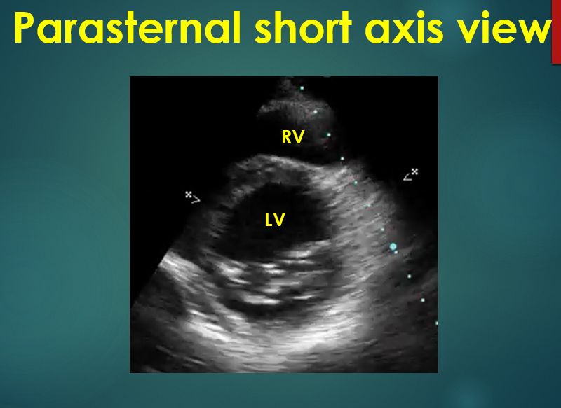
Parasternal short axis view is obtained by rotating the transducer almost at right angle in the same location so that the echo beam is perpendicular to the base apex axis of the heart. Three cuts are usually obtained in this view. The aorta, right ventricular outflow tract and pulmonary artery up to its bifurcation is imaged in the upward angulation shown in the left panel. Colour flow shows the flow in pulmonary artery.
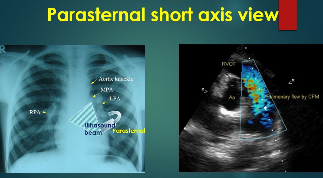
Slight downward angulation of the transducer from this view gives the left ventricular cross section with mitral valve cross section within. Right ventricular cavity is elliptical in this view and left ventricular cavity is circular. Wall motion of the left ventricle can be assessed in this view also. Planimetry of mitral valve area can be obtained in parasternal short axis view in case of mitral stenosis.
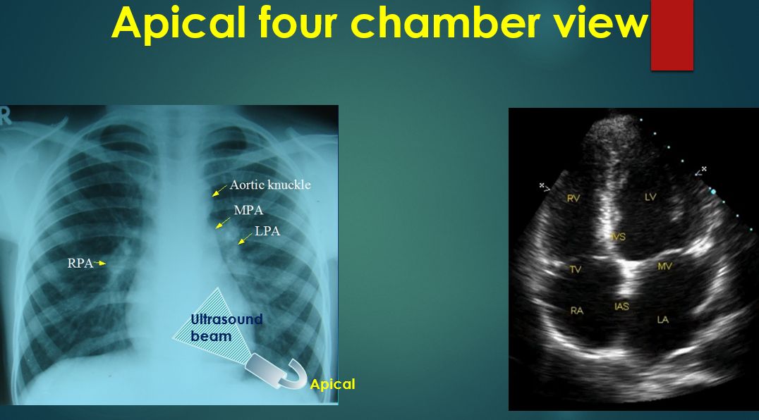
Apical views are obtained by keeping the transducer directly over the apex beat. Apical four chamber, two chamber and three chamber views can be obtained by rotating the transducer. Apical four chamber view is illustrated here. Apical four chamber view shows all four cardiac chambers, mitral and tricuspid valves, and the septa. But as the echo beam is parallel to the interatrial septum, false echo drop outs can occur in the interatrial septum. Apical five chamber view including the proximal aorta can be obtained by slight tilting of the transducer from this view.
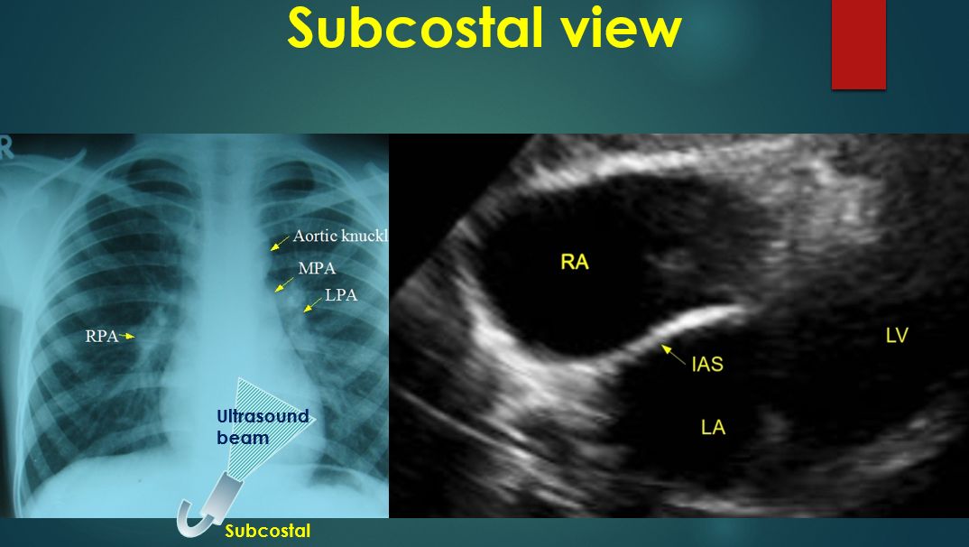
Subcostal view is obtained from below the xiphisternum in the epigastrium. Transducer rotation is needed to get subcostal four chamber and short axis views. Interatrial septum is best imaged in this view. Assessment of the inferior vena cava for checking the hydration status is also feasible from this view. Subcostal view is a favourite view of pediatric echocardiographers.
Suprasternal view is useful in imaging arch of the aorta and nearby regions of ascending and descending aorta. Coarctation of aorta and patent ductus arteriosus can be imaged in this view. The image shows the blue coloured descending aortic flow on colour Doppler. Gradient of coarctation can be assessed in this view. Apart from these standard views, other modified views may also be used in certain circumstances. A right parasternal view may be used when the heart is enlarged, to assess the tricuspid regurgitation jet. Dilated ascending aorta will also be visible in a right parasternal view.
Related Posts

About The Author
Johnson Francis
Former Professor of Cardiology, Calicut Govt. Medical Kozhikode, Kerala, India. Editor-in-Chief, BMH Medical Journal