Pericardial effusion on echo
Mild pericardial effusion on echocardiography
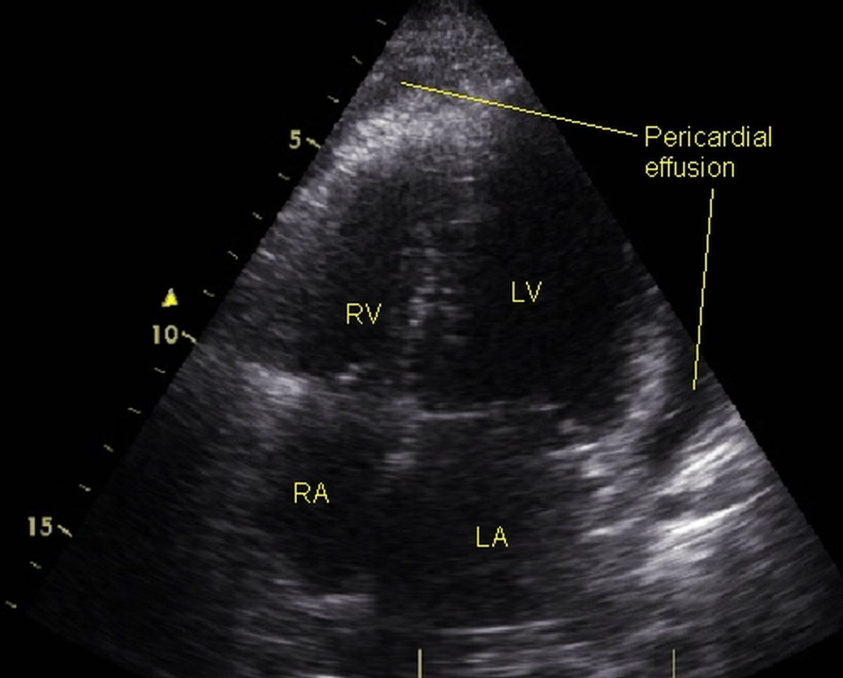 Mild pericardial effusion on echocardiography from apical four chamber view
Echocardiogram from the apical four chamber view shows mild pericardial effusion as echo free space lateral to the left ventricle and anteromedial to the right ventricle. The quantity of effusion is small and needs only observation. It will be difficult to aspirate such small effusions without injuring the cardiac structures. RV: right ventricle; RA: right atrium; LV: left ventricle; LA: left atrium. Pericardial fat can produce echo free space and can be mistaken for pericardial effusion on echocardiography, but can be differentiated by magnetic resonance imaging.
The usual route for pericardial aspiration is subcostal. Hence it is always better to check the size of pericardial effusion from subcostal view. Aspiration is usually done only if there is a sizable pericardial effusion anterior to the right ventricle in the subcostal view. If the effusion is small, the aspiration needle is likely to injure the right ventricle. Pigtail catheter is often introduced into the pericardial space using Seldinger technique with guidewire and left there till the pericardial effusion stops collecting. This method is especially useful in malignant pericardial effusion in which repeated aspirations are likely to be necessary. In such situations, sclerosing agents like Bleomycin can be instilled into the pericardial space after complete aspiration, to cause adhesion of pericardial layers (visceral and parietal) and prevention of recurrence.
Quantification of pericardial effusion
In general pericardial effusion less than 10 mm can be considered mild, 10-20 mm moderate and more than 20 mm as large. There have been attempts to quantify pericardial effusion by echocardiography. In one report, they could get good correlation between echocardiographically measured volume and surgically drained volume of pericardial effusion in 20 patients with 100 to 1200 ml of pericardial fluid [1].
Reference
- Prakash AM, Sun Y, Chiaramida SA, Wu J, Lucariello RJ. Quantitative assessment of pericardial effusion volume by two-dimensional echocardiography. J Am Soc Echocardiogr. 2003 Feb;16(2):147-53.
Related Posts


About The Author
Johnson Francis
Former Professor of Cardiology, Calicut Govt. Medical Kozhikode, Kerala, India. Editor-in-Chief, BMH Medical Journal