Cardiomegaly due to LV dysfunction on X-ray chest PA view
Cardiomegaly due to LV dysfunction on X-ray chest PA view
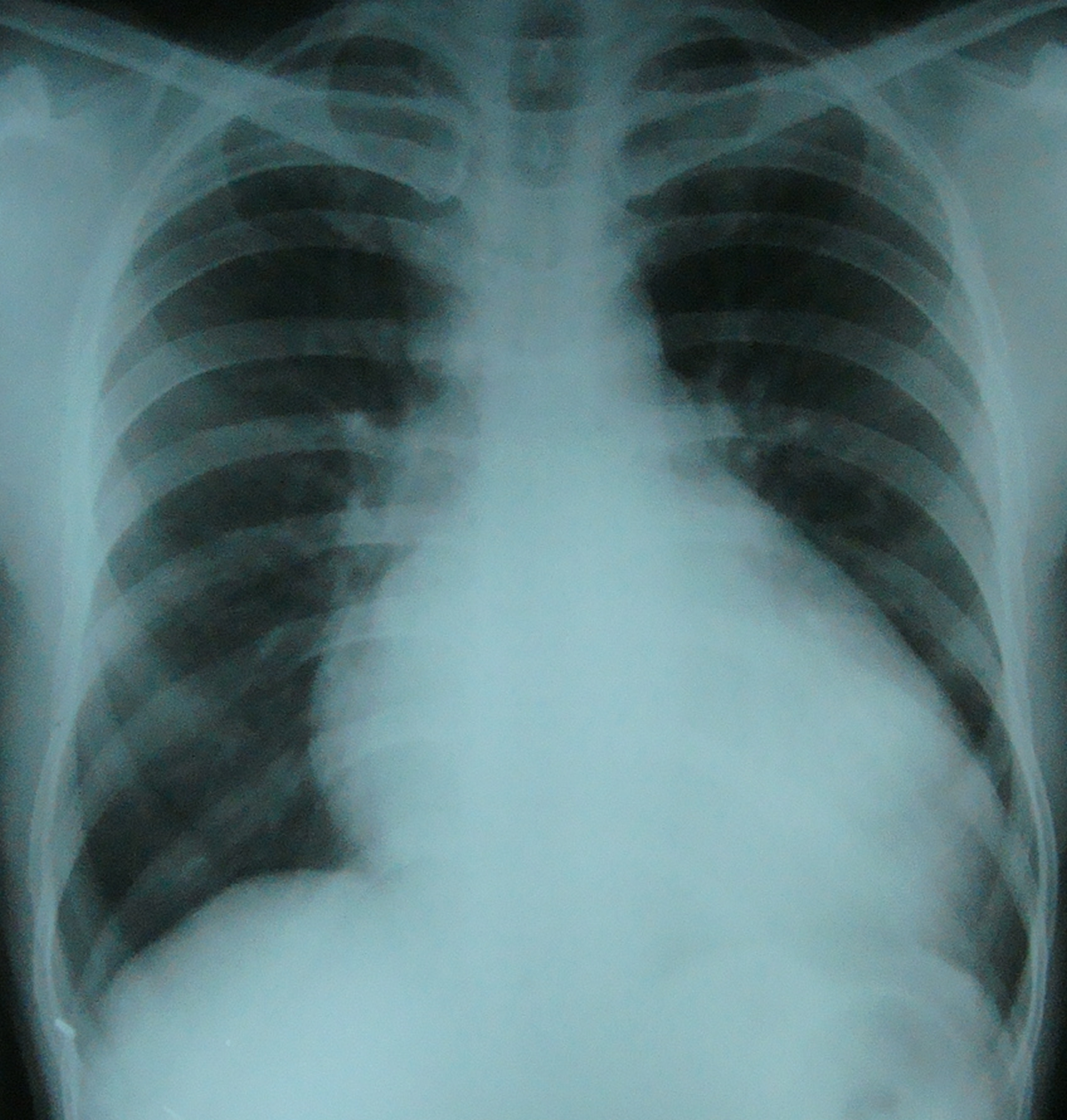
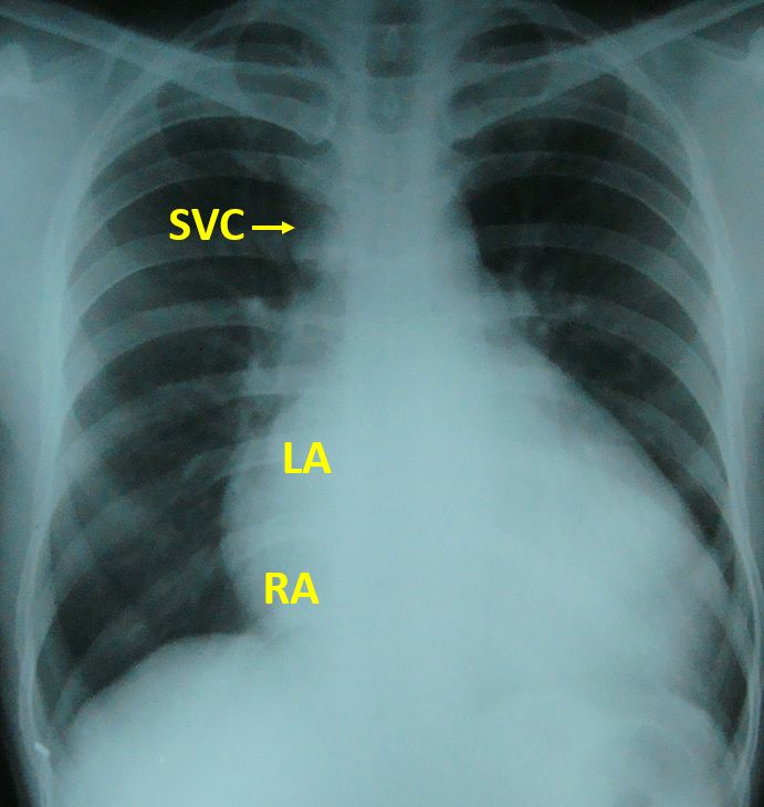
X-ray chest PA view showing cardiomegaly due to left ventricular (LV) dysfunction. Right border is seen significantly enlarged rightwards indicating right atrial enlargement. A shadow within shadow is also seen near the right border indicating left atrial enlargement. A superior vena caval shadow is extending up from the right cardiac border. Clinical presentation was with exertional dyspnoea. Cardiomegaly was clinically evident and there was a left parasternal heave indicating right ventricular enlargement due to pulmonary arterial hypertension which was secondary to left ventricular dysfunction and elevated pulmonary venous pressures.
Electrocardiogram showed left atrial enlargement as a sequelae of left ventricular dysfunction, poor progression of R waves in anterior leads indicating old anterior wall myocardial infarction and T wave inversions in lateral leads.
Related Posts


About The Author
Johnson Francis
Former Professor of Cardiology, Calicut Govt. Medical Kozhikode, Kerala, India. Editor-in-Chief, BMH Medical Journal