Simultaneous LV and RV angios (left ventricular and right ventricular angiograms or ventriculograms) used to be done in hypertrophic cardiomyopathy (asymmetric septal hypertrophy) before the advent of echocardiography to delineate the
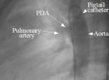
Serial angiographic shots showing steps in device closure of patent ductus arteriosus (PDA). Though small PDAs can be closed by coil occlusion, larger ones require device closure.
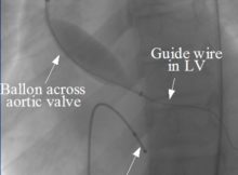
Balloon aortic valvotomy (BAV) is usually done through the transfemoral route. Potential risks include balloon rupture and embolisation of calcific material, apart from access site issues.
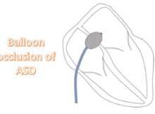
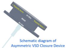
Deficiency of aortic rim an important risk factor for cardiac erosion with ASD device Cardiac erosion is a potential nightmare of device closure of atrial septal defect (ASD)
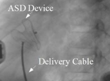
Pulmonary edema after device closure of ASD Device closure of atrial septal defect (ASD) is almost replacing surgical ASD closure in all cases with suitable morphology. Percutaneous ASD
Transcatheter repair of tricuspid valve Transcatheter repair of tricuspid valve: An article in JACC [1] has highlighted the possibility of transcatheter repair of tricuspid valve in severe tricuspid
Coil embolization of Patent Ductus Arteriosus (PDA) is one of the options for non-surgical closure of ductus. The other non-surgical option is device closure with Amplatzer PDA occluder.