Conventionally, aortic stenosis has been classified into mild (peak gradient up to 50 mm Hg), moderate (peak gradient between 50 - 75 mm Hg) and severe, with
Transmitral Doppler tracing from apical four chamber view showing varying degrees of fusion of mitral E and A waves. E/A reversal is also evident in the later three
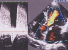
Pressure half time of aortic regurgitation jet is measured from the apical five chamber view using continuous wave Doppler echocardiography. Pressure half time decreases as the severity of
Echocardiogram video showing large aortic valve vegetation and prolapse of a scallop of the anterior mitral leaflet. Left atrium and left ventricle are dilated due to mitral and