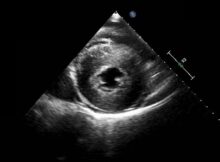
LVH on echocardiogram Parasternal short axis view at the level of the papillary muscles showing severe concentric left ventricular hypertrophy. Serial short axis views have to be obtained
D shaped LV cavity in severe RVH Echo from parasternal short axis view shows a D shaped LV cavity (left ventricular cavity) alongside a grossly hypertrophied right ventricle
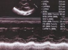
M-Mode echocardiogram at Aorta – Left atrium level M-Mode echocardiogram at Aorta – Left atrium level: M-Mode cuts are usually obtained from two dimensional (2-D) views after the proper
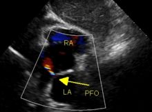
Atrial septal defect with right to left shunt should prompt us to look for total anomalous pulmonary venous connection as well as severe pulmonary hypertension.
M-Mode echocardiography and Anatomical M-Mode M-Mode echocardiography (Time-motion mode) was one of the earliest tools of the echocardiographer. M-Mode gives an ice-pick view of the heart. The vertical
Left atrial myxoma – echocardiogram with video in parasternal long axis view with systolic and diastolic frames. Mitral regurgitation is also seen on color Doppler.
Aorta – left atrium M-mode Mode echocardiogram at aorta-left atrium level Mode tracing at the aorta-left atrium level is often the first M-mode tracing to be taken from the
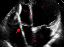
Patent foramen ovale – PFO Patent foramen ovale is a valvular opening in the fossa ovalis region of the interatrial septum. In fetal life, the foramen ovale shunts
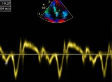
Diastolic dysfunction by tissue Doppler E/E’ measurement by Tissue Doppler Imaging E/E’ measurement is used to assess diastolic function by tissue Doppler. Method may vary in technical details