Atrial septal defect (ASD) with bidirectional shunt Atrial septal defect (ASD) with bidirectional shunt: Subcostal four chamber view shows a large defect in the interatrial septum (ASD). Right
Severe tricuspid regurgitation – echocardiogram Echocardiogram in apical four chamber view shows severe tricuspid regurgitation as a large mosaic jet filling more than half of a dilated right
Monomorphic ventricular premature complex (VPC) Basic rhythm in the ECG is sinus rhythm at around 60/min. There are two wide QRS complexes seen in lead II rhythm strip.
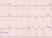
Old inferior wall infarction and lateral ST depression ECG shows sinus rhythm at around 75/min, with pathological Q waves in inferior leads, indicating old inferior wall myocardial infarction.
Popliteal angiogram Popliteal angiogram is usually obtained as part of the femoral angiogram by panning the table downwards to cover the popliteal artery as the contrast flows down
Evolved anterior wall myocardial infarction ECG shows sinus rhythm at a rate of around 100/min, with QS complexes in anterior leads along with a coved ST segment elevation
Pacing in complete heart block – ECG Pacing in complete heart block (CHB): Initial part of the ECG shows narrow QRS complexes at a rate of around 43/minute.