CXR showing dilated RVOT Chest X-ray PA view showing a bulge along the left border, below the aortic knuckle and pulmonary bay. Sternal wires are seen near the mid line, which would
Risk factor/s for upper limb venous thrombosis: a) Vigorous arm exercise b) Central venous catheter c) Pacemaker / defibrillator leads d) All of the above Correct answer: d)
Wrong statement about deep vein thrombosis of upper limbs compared to lower limbs: a) Lower chance of pulmonary embolism b) Higher recurrence at one year c) Lower incidence
Risk of intracranial bleed for tenecteplase in acute pulmonary embolism: a) 2% b) 5% c) 10% d) 15% Correct answer: a) 2% The incidence hemorrhagic stroke after thrombolysis
Surgical embolectomy in pulmonary embolism is indicated in: a) Impending paradoxical systemic embolism b) Hypotension with contraindication for thrombolysis c) Failed thrombolysis d) All of the above Correct
True regarding inferior vena caval (IVC) filter for prevention of pulmonary embolism: a) Indicated when anticoagulation is contra indicated b) Recurrence in spite of anticoagulation c) Increases risk
Drugs approved for treatment of normotensive patients with pulmonary embolism: Heparin Fondaparinux Rivaroxaban Apixaban Dabigatran Edoxaban Thrombolytic therapy is reserved for rescue in case of decompensation due to
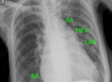
Chest X-ray in Mitral Stenosis with third mogul sign Main pulmonary artery segment is usually concave on a normal chest X-Ray. Here it is seen as convex, bulging