Coronary venous circulation
Coronary venous circulation
Importance of knowing the coronary venous circulation has increased in recent years with use of cardiac resynchronization therapy (CRT) for heart failure. Left ventricular epicardial pacing in CRT is achieved by placing a lead in a coronary vein, usually a posterolateral tributary of the coronary sinus.
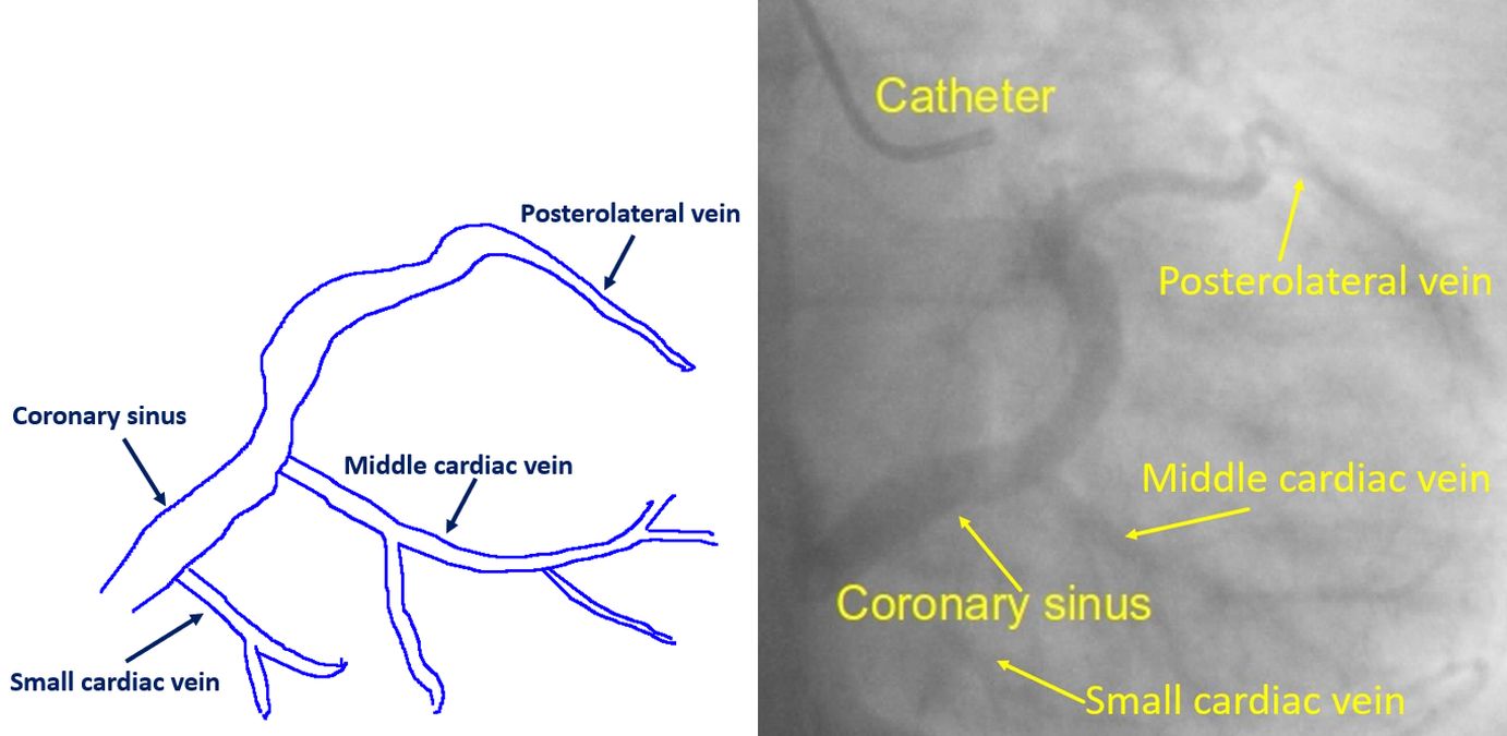
Coronary venous circulation can be visualized by a coronary angiogram levophase. After filling the coronary arteries, the contrast drains into the coronary venous system, finally opacifying the coronary sinus, which drains into the right atrium. Here is a coronary angiogram levophase, with a line diagram beside it:
To get better visualization of the coronary sinus and its tributaries, a balloon occlusion angiography of the coronary sinus is done by cannulating it retrogradely from the right atrium. But that is done only just prior to CRT as there is a small risk of coronary sinus dissection. The veins visualized in a coronary angiogram levophase will also depend on the coronary artery in which contrast is injected.
Coronary venous circulation has been divided into a greater cardiac venous system which drains into the coronary sinus and a smaller cardiac venous system which drains into cardiac chambers. Smaller cardiac venous system is constituted mainly by the Thebesian veins [1].
Coronary sinus is the largest cardiac vein with multiple tributaries. It is located in the left posterior atrioventricular groove and empties directly into the right atrium through the coronary sinus ostium. Tributaries of the coronary sinus are the great cardiac vein, middle cardiac vein, small cardiac vein, left posterior left ventricular vein and oblique cardiac vein.
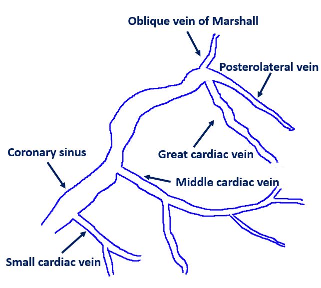
Great cardiac vein starts from the apex and drains upwards, in the anterior interventricular sulcus. In the initial part, it is also known as anterior interventricular vein. In the anterior interventricular groove it lies alongside the left anterior descending coronary artery. At the base of the heart it travels leftwards and curves posteriorly near the left atrial appendage. Posteriorly it converges with the oblique vein of left atrium (oblique vein of Marshall) and forms the coronary sinus. Oblique vein of Marshall is the residua of the embryonic left superior cardinal vein [2]. Posterolateral vein is directly opposite the vein of Marshall.
Middle cardiac vein also originates at the apex, but drains the posterior side of the heart and is situated in the posterior interventricular sulcus and empties into the coronary sinus. It runs along with the posterior descending coronary artery, but in reverse direction. Small cardiac vein is in the posterior part of right atrioventricular sulcus. It also drains into the coronary sinus.
Valve at the ostium of the coronary sinus is known as Thebesian valve. Valve at the ostium of the posterolateral vein at the junction of the great cardiac vein and coronary sinus is called Vieussen’s valve [3]. These valves can cause difficulties during ablation and CRT procedures.
Another practical importance of the coronary venous system is that oblique vein of Marshall can give rise to atrial fibrillation. Three reasons described are the myocardial extensions into the structure, node like remnants within the vein and the rich autonomic innervation surrounding it [3].
References
- Miao JH, Makaryus AN. Anatomy, Thorax, Heart Veins. [Updated 2020 Jul 31]. In: StatPearls [Internet]. Treasure Island (FL): StatPearls Publishing; 2021 Jan-. Available from: https://www.ncbi.nlm.nih.gov/books/NBK549786/
- Harikrishnan S, Nair K, Tharakan J. Oblique vein of Marshall. Heart. 2005 Feb;91(2):e16.
- Habib A, Lachman N, Christensen KN, Asirvatham SJ. The anatomy of the coronary sinus venous system for the cardiac electrophysiologist. Europace. 2009 Nov;11 Suppl 5:v15-21.
Related Posts

About The Author
Johnson Francis
Former Professor of Cardiology, Calicut Govt. Medical Kozhikode, Kerala, India. Editor-in-Chief, BMH Medical Journal