ECG Quiz 59 – Discussion
ECG Quiz 59 – Discussion
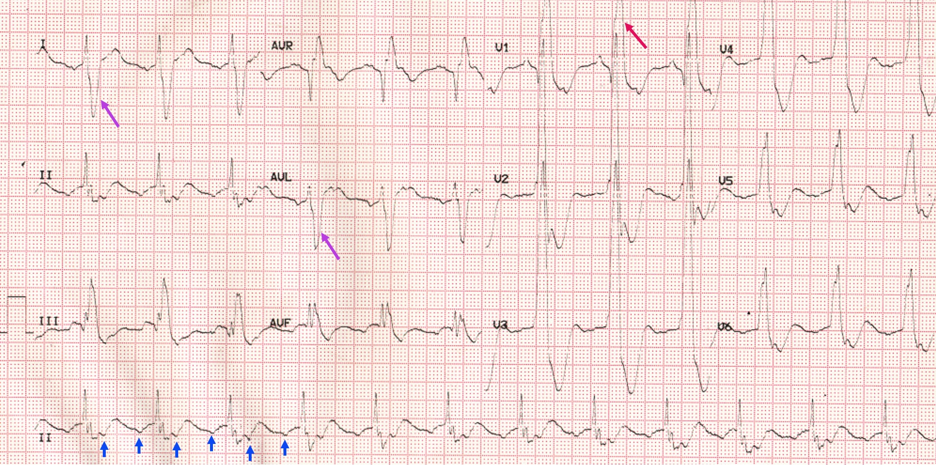
P waves can be seen at a rate double that of QRS complexes (blue arrows). They are inverted in inferior leads, indicating an ectopic focus from the low atrium (ectopic atrial tachycardia). 2:1 conduction excludes the possibility of an AV node mediated tachycardia which would get terminated with the onset of AV conduction block.
The QRS is very wide (200 ms), with a tall slurred R’ in V1 and a slurred S wave in leads I and aVL. These indicate a complete right bundle branch block. QRS has right axis deviation suggestive of left posterior hemiblock in addition. Fragmented QRS is visible in inferior leads (multiple notches in QRS complex). All these indicate a high risk for ventricular arrhythmias. Such wide QRS is often noted in post operative cases of Tetralogy of Fallot.
About The Author
Johnson Francis
Former Professor of Cardiology, Calicut Govt. Medical Kozhikode, Kerala, India. Editor-in-Chief, BMH Medical Journal