ECG showing atrial and ventricular pacing spikes
ECG showing atrial and ventricular pacing spikes
(Click on the image for an enlarged view)
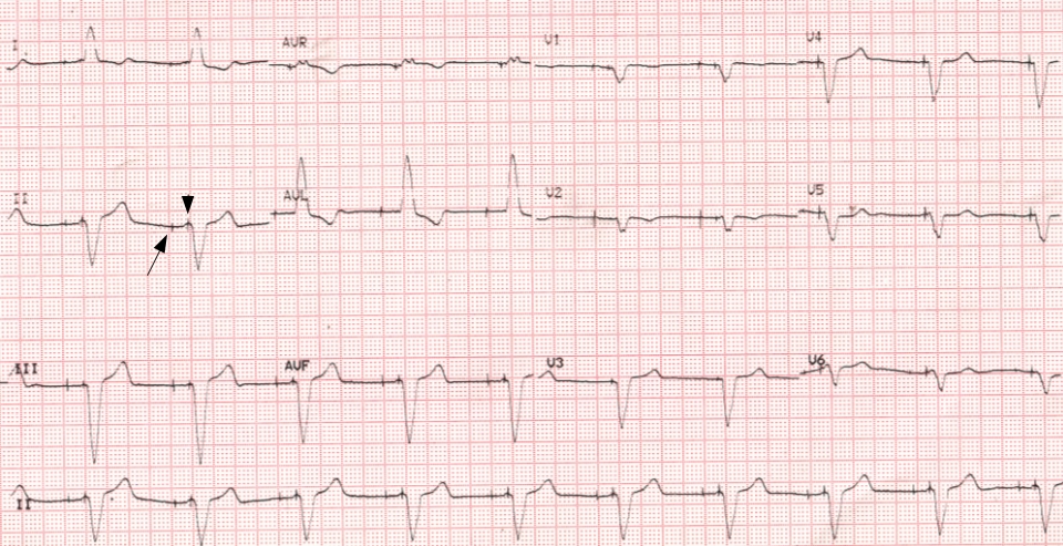
ECG showing atrial (long arrow) and ventricular (short arrow) pacing spikes, also known as pacing artifacts. Atrial pacing spikes precede the ventricular pacing spike by the programmed AV interval. In this ECG, though the atrial pacing spikes are seen, atrial capture in the form of paced P waves are not visible. It is possible that there is atrial non-capture either due to high thresholds or an atrial lead displacement. Another possibility is that paced P waves are isoelectric in the recorded leads.
Regular ventricular capture is seen after each ventricular spike. The paced QRS complexes are wide and having left bundle branch block pattern with left axis deviation suggesting a ventricular pacing site at right ventricular apex (right ventricular apical pacing). Similar pattern with normal QRS axis would suggest pacing from the right ventricular outflow region (RVOT pacing).
Pacing artifacts are high frequency signals picked up from the pulse delivered by the pacemaker for electrical stimulation of a cardiac chamber. It is usually well documented by an analog ECG (electrocardiograph) machine. When it comes to digital ECG machines, the pacing artifacts may be ironed out by the low pass filter settings which do not permit the display of the high frequency pacing signal. Setting the upper limit of the frequency response (low pass filter) to around 100 Hz usually displays the pacing artefact well.
Some authors have even recommended using a low pass filter of 300 Hz to improve detection of pacemaker spike [1]. They studied low pass filter settings from 40 Hz to 400 Hz in 12 lead ECG. Among the low pass filter settings studied, 300 Hz had the best performance in detecting atrial pacing spikes. The gold standard for comparison was the diagnosis of pacemaker programmer.
Reference
- Jian Sun, Qiu-Feng Lu, Yan Zhao, Peng-Pai Zhang, Jun Wang, Qun-Shan Wang, Xiao-Hong Liu, Yi-Gang Li. A low-pass filter of 300 Hz improved the detection of pacemaker spike on remote and bedside electrocardiogram. Chin Med J (Engl). 2019 Mar 5;132(5):534-541.
Related Posts

About The Author
Johnson Francis
Former Professor of Cardiology, Calicut Govt. Medical Kozhikode, Kerala, India. Editor-in-Chief, BMH Medical Journal