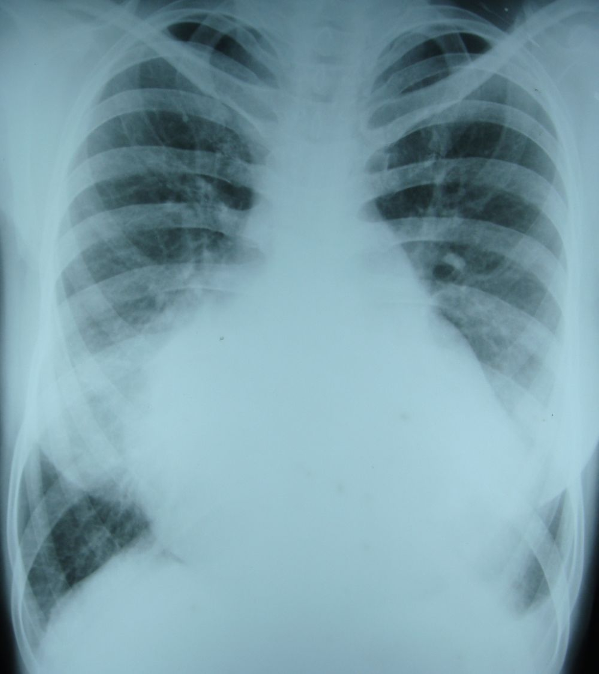
X-ray chest PA view in biventricular endomyocardial fibrosis
X-ray chest PA view in biventricular endomyocardial fibrosis
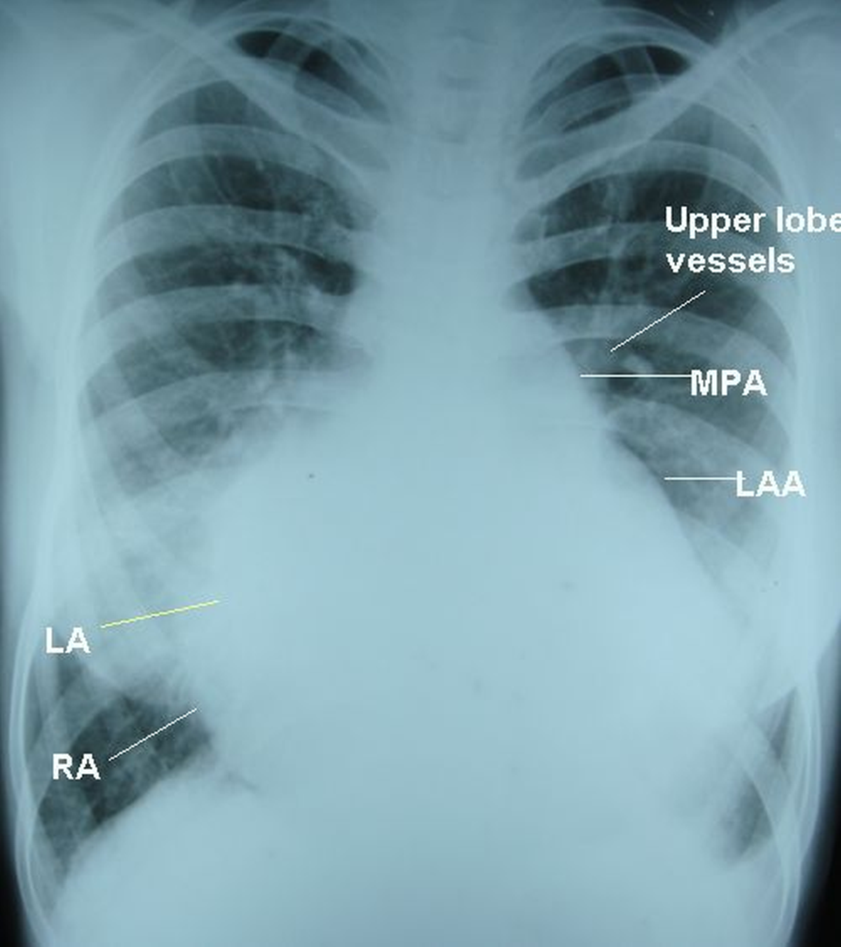
(See annotated image below)
X-ray chest PA view in biventricular endomyocardial fibrosis. There is cardiomegaly with right atrial enlargement (shift of the right border of the heart to the right). A double atrial shadow (shadow in shadow) is seen within the right atrial contour, suggesting left atrial enlargement. Left atrial appendage and main pulmonary artery segment are prominent on the left upper cardiac border. The upper lobe vessels are dilated, indicating pulmonary venous hypertension. The differential diagnosis of this x-ray would be mitral stenosis with pulmonary hypertension ± tricuspid valve disease. Right cardiophrenic shows a few horizontal lines suggestive of Kerley B lines. Left cardiophrenic angle is obliterated by a small pleural effusion (compare with right cardiophrenic angle).
Related Posts

About The Author
Johnson Francis
Former Professor of Cardiology, Calicut Govt. Medical Kozhikode, Kerala, India. Editor-in-Chief, BMH Medical Journal