Initiation and spontaneous termination of SVT
Initiation and spontaneous termination of SVT
Initial monitor screen shot shows initiation of SVT and the second one shows spontaneous termination of SVT.
SVPC to SVT
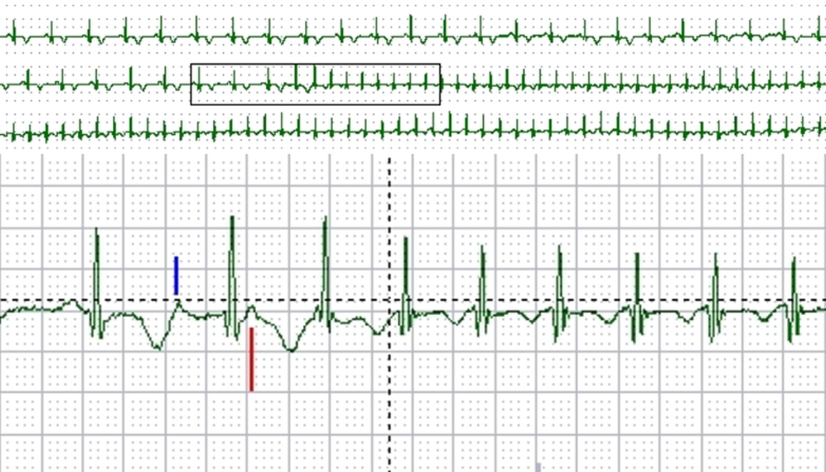
Monitor screenshot showing initiation of SVT by an SVPC
SVT: Supraventricular tachycardia; SVPC: Supraventricular premature complex
Sinus rhythm with normal PR interval is seen in the initial beats. A supraventricular ectopic (blue marker) is conducted with long PR interval as the fast pathway (AV nodal) has not recovered fully after conduction of the previous sinus beat. This leads to an echo beat (red marker). Supraventricular tachycardia (SVT) can be seen subsequently. The sudden increase in PR interval with a supraventricular ectopic beat is equivalent to the PR jump noted during EP (electrophysiology) study and indicates dual AV nodal physiology. PR jump of 50 milliseconds or more is considered indicative of dual AV nodal physiology.
SVT to SR
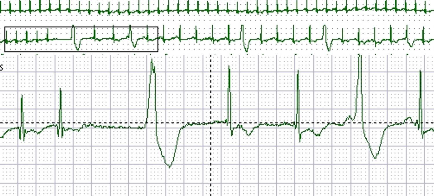
Monitor screenshot showing spontaneous termination of SVT to sinus rhythm (SR).
Initial 2 beats are part of the supraventricular tachycardia. Termination of tachycardia leads to a short pause and ventricular escape beat (wide QRS). This is followed by two narrow QRS beats, the P waves of which are not well seen (possibly of low amplitude). Next wide QRS complex is preceded by a P wave, indicating a late diastolic ventricular ectopic beat. The QRS width of this beat is lesser than that of the previous ventricular escape beat. Finally there is a good amplitude P wave followed by a narrow QRS after a normal PR interval, indicating sinus rhythm (SR).
Related Posts


About The Author
Johnson Francis
Former Professor of Cardiology, Calicut Govt. Medical Kozhikode, Kerala, India. Editor-in-Chief, BMH Medical Journal