Left ventricular aneurysm – X-ray chest PA
Left ventricular aneurysm – X-ray chest PA
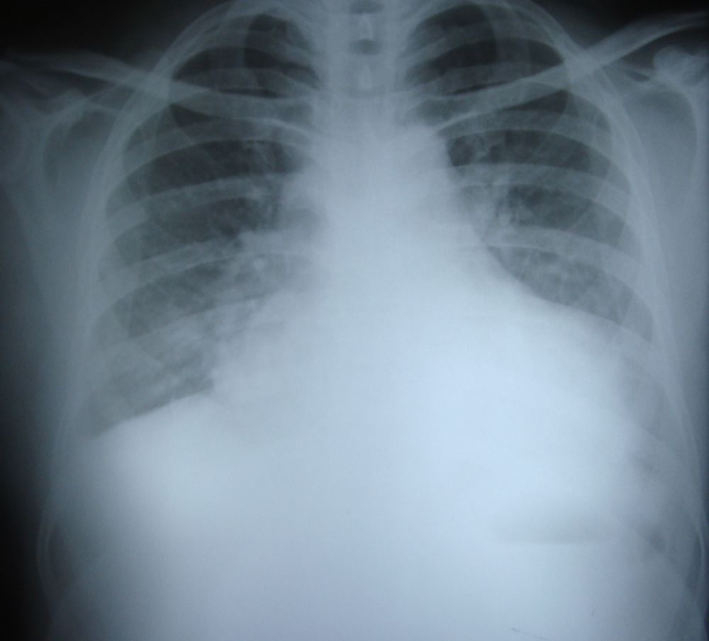
The bulge along the left border in this X-ray chest PA view is due to a left ventricular aneurysm. There is associated pulmonary congestion due to heart failure which often accompanies left ventricular aneurysm due to wasted systolic effort of the left ventricle. The aneurysm bulges out in systole and part of the left ventricular effort is lost in expanding the aneurysm, instead of ejecting the blood into the aorta. This aneurysm was secondary to anterior wall myocardial infarction. Clinically there was a dyskinetic see-saw pulsation in the apical region. Echocardiogram confirmed the presence of a true left ventricular aneurysm. A true aneurysm has all three layers of the heart: pericardium, myocardium and endocardium, while a false aneurysm is a contained rupture bounded only by the pericardium. A true aneurysm can cause heart failure and ventricular arrhythmias, but seldom ruptures, while a false aneurysm has a high chance of secondary rupture. True aneurysm usually has a wide neck while a false aneurysm has a narrow neck.
Related Posts

About The Author
Johnson Francis
Former Professor of Cardiology, Calicut Govt. Medical Kozhikode, Kerala, India. Editor-in-Chief, BMH Medical Journal