LVEMF with calcification -echocardiograms
LVEMF with calcification -echocardiograms
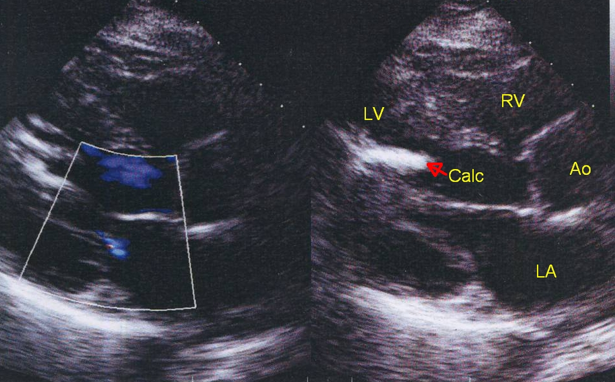
Echocardiogram in parasternal long axis (PLAX) view demonstrating a region of dense calcification (Calc) in the left ventricle, in a case of left ventricular endomyocardial fibrosis. LV: left ventricle; RV: right ventricle; Ao: aorta; LA: left atrium.
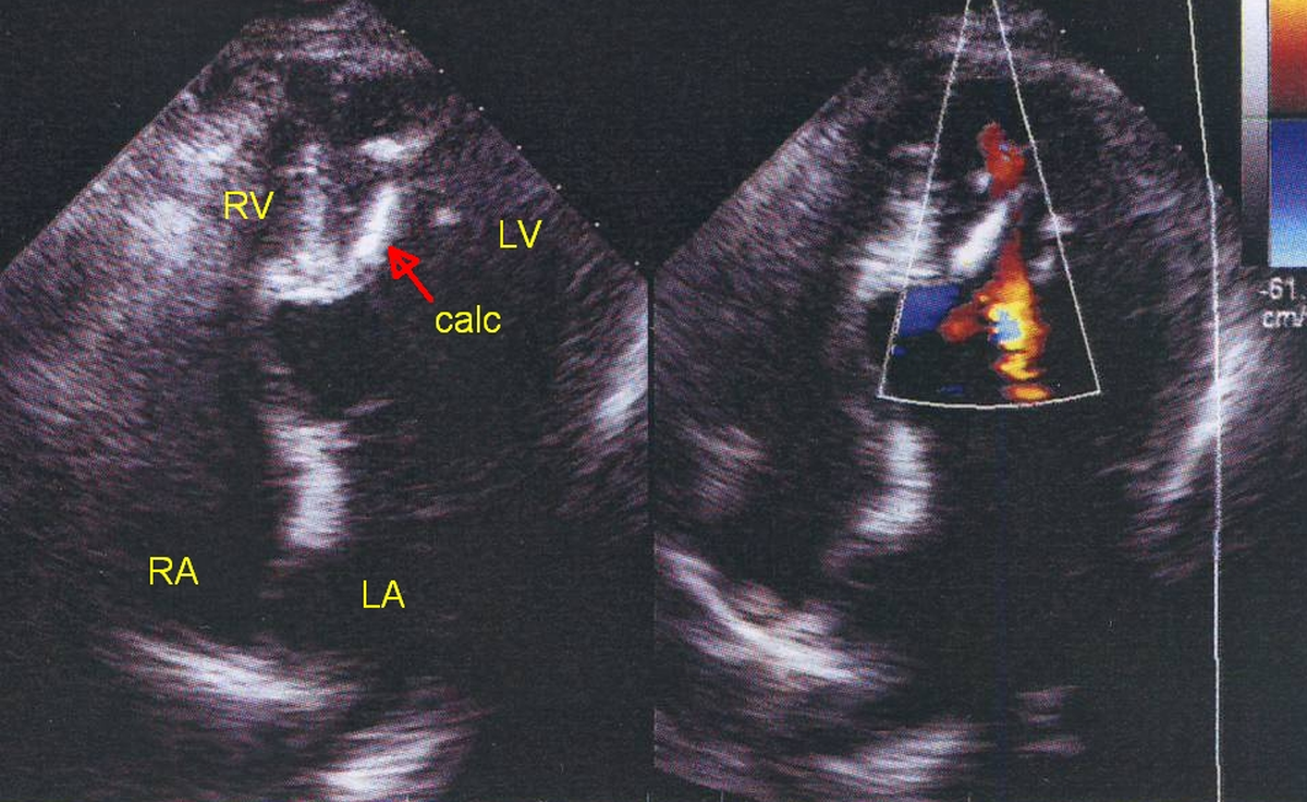
Echocardiogram in apical four chamber (4C) view demonstrating a region of dense calcification (Calc) in the left ventricle, in a case of LVEMF. RA: right atrium
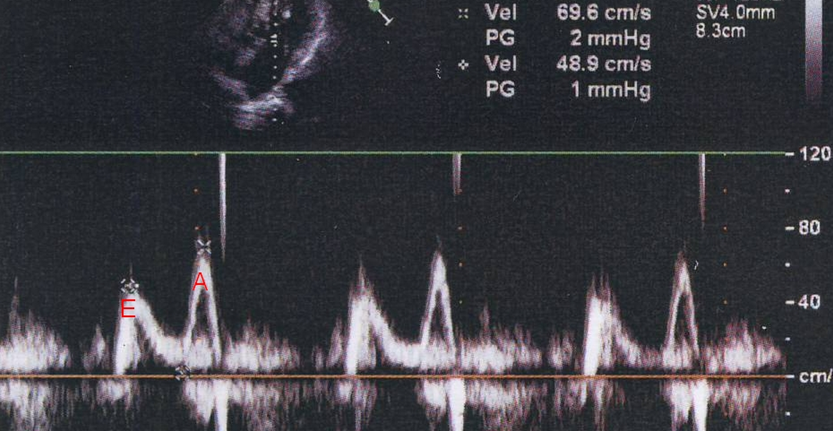
E wave is taller than A wave in the mitral inflow tracing, suggesting left ventricular diastolic dysfunction.
Calcification can also occur due to persistently elevated serum calcium levels as in primary hyperparathyroidism, chronic kidney disease, hypervitaminosis D and extensive bone destruction due to metastatic disease or multiple myeloma.
Dystrophic calcification without persistent hypercalcemia can occur in ischemic heart disease, primary myocardial disease and Loeffler endocarditis due to prolonged eosinophilia.
Reference
- Khanna R, Kapoor A, Soni N. A Heart Set in Stone: A Case of Extensive Cardiac Calcification. Heart Views. 2016 Jul-Sep;17(3):100-102.
About The Author
Johnson Francis
Former Professor of Cardiology, Calicut Govt. Medical Kozhikode, Kerala, India. Editor-in-Chief, BMH Medical Journal