Low atrial rhythm – ECG
Low atrial rhythm
Abstract: Low atrial rhythm manifests with inverted P waves in inferior leads. It may be seen in sinus venosus atrial septal defect.
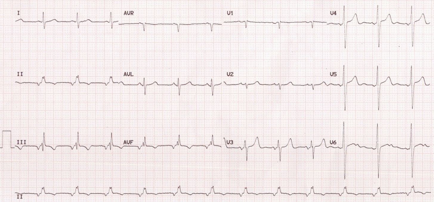
This ECG shows inverted P waves in inferior leads (II, III and aVF). This indicates that the atrial activation is spreading from below upwards. It is suggestive of a focus either in the low atrium or high junction. A mid junctional rhythm will have no visible P waves as the P wave will be within the QRS due to simultaneous activation of the atria and ventricles. In low junctional rhythm the P wave occurs after the QRS, in the ST segment and is inverted in inferior leads. In left atrial rhythm originating from the lower part, the P waves are inverted in inferior leads as well as lateral leads.
There is notching of the QRS complex in the inferior leads which suggest the crochetage sign in atrial septal defect. This rhythm can occur with sinus venosus atrial septal defect as the sinus node may be defective so that alternate focus arising in the low atrium gives the dominant rhythm. The PR interval is also shorter in low junctional and low atrial foci, more in the former than in the latter, due to obvious reasons.
It has been reported in acute amlodipine intoxication [1]. A rare autosomal dominant disorder in four generations of a family with congenital heart diseases (atrial septal defect, tetralogy of Fallot and persistent left superior vena cava) and low atrial rhythm has also been documented recently [2].
An interesting case report in which low atrial rhythm mimicked myocardial infarction has been reported recently [3]. In their case, the person presented with left forearm pain and numbness. Coronary angiography was normal even though ECG showed low atrial rhythm and inferior ST segment elevation. Numbness and pain of the left arm was eventually shown to be due to carpal tunnel syndrome. Authors had a novel explanation for the ST segment elevation as being due to atrial repolarization wave (Ta wave), which can extend up to the ST segment in faster rhythm. It is known that Ta wave can cause ST depression during treadmill exercise test. In low atrial rhythm, as the atrial depolarization proceeds from below upwards, atrial repolarization wave can proceed in the opposite direction and cause ST segment elevation in inferior leads.
References
- Miranda CH, Xavier L, Fiorante F, Misiara GP, Guimarães EG, Galli AM, Pazin-Filho A, de Carvalho Borges M. Cardiac rhythm disturbances associated with amlodipine acute intoxication. Cardiovasc Toxicol. 2012 Dec;12(4):359-62.
- van de Meerakker JB, van Engelen K, Mathijssen IB, Lekanne dit Deprez RH, Lam J, Wilde AA, Baars MJ, Mannens MM, Mulder BJ, Moorman AF, Postma AV. Eur J Hum Genet. 2011 Jul;19(7):820-6. (Free Full Text at Pubmed Central).
- Arı H, Kahraman F, Baş HA, Arslan A. Anatol J Cardiol. 2015 Aug;15(8):675, 683. (Free Full Text at Pubmed Central)
Related Posts

About The Author
Johnson Francis
Former Professor of Cardiology, Calicut Govt. Medical Kozhikode, Kerala, India. Editor-in-Chief, BMH Medical Journal