Mitral Gradient by CW Doppler in Mitral Stenosis
Mitral gradient by CW Doppler in mitral stenosis
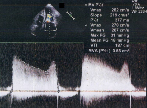
Mitral gradient by CW Doppler in mitral stenosis demonstrating the maximum gradient of 31 mm Hg and mean gradient of 18 mm Hg. The Doppler tracing is obtained by aligning the Doppler cursor along the trans mitral jet during diastole in the apical four chamber view. The outline of the tracing is sketched out manually to measure the velocities and gradient, which is calculated by the software package in the echocardiograph. Pressure half time of the initial slope is calculated as 377 milliseconds. VTI: velocity time integral; MVA: mitral valve area. Here the mitral valve area calculated by the pressure half time method is 0.58 square cm, indicating critical mitral stenosis.
The large left atrium seen in the upper panel is also consistent with severe mitral stenosis. Vmax of 282 cm/s is the peak velocity of the initial peak and the Vmax of 278 cm/s is the peak velocity of the second peak. Even though the A wave is blunted in mitral stenosis in the M-mode echocardiogram, the A of Doppler tracing is not. Slope of 219 cm/s is the slope following the initial E, which is used to estimate mitral valve area. Pressure half time is calculated from the peak velocity and the slope by the software package. The mitral valve area is derived from the pressure half time by dividing 220 by pressure half time. This is because a pressure half time of 220 ms corresponds to a mitral valve area of 1 square cm. Continuous wave Doppler is needed because the velocities are beyond the usual range for pulsed Doppler. If pulsed Doppler is used, the signals will get aliased as the velocity is beyond the Nyquist limit of the pulsed Doppler system.
Related Posts

About The Author
Johnson Francis
Former Professor of Cardiology, Calicut Govt. Medical Kozhikode, Kerala, India. Editor-in-Chief, BMH Medical Journal