Mitral Valve Prolapse (MVP) and Sudden Cardiac Arrest
While mitral valve prolapse (MVP) is generally a benign condition with an excellent overall prognosis, a specific, albeit rare, subset of patients exhibit what is termed the Arrhythmic Mitral Valve Prolapse (AMVP) or “Malignant MVP” phenotype. These patients are at a distinctly elevated risk for complex ventricular arrhythmias and sudden cardiac arrest (SCA). The 2022 EHRA expert consensus statement provides the current framework for identifying and managing this high-risk cohort.
The Pathophysiologic Mechanism
The link between MVP and arrhythmogenesis is primarily mechanical, leading to an acquired structural substrate.
- Mechanical Stretch: The severe systolic billowing of myxomatous leaflets exerts excessive traction on the chordae tendineae and the papillary muscles.
- Myocardial Hypertrophy and Fibrosis: This chronic, localized mechanical stress leads to friction and stretch on the inferobasal left ventricular wall and papillary muscles, causing localized myocardial hypertrophy and, eventually, replacement fibrosis.
- Arrhythmogenic Focus: This fibrotic substrate alters local conduction properties, creating a nidus for re-entry and triggering premature ventricular contractions (PVCs). These PVCs can initiate polymorphic ventricular tachycardia (VT) or ventricular fibrillation (VF), culminating in SCA.
Identifying the “Malignant” Phenotype
Risk stratification relies on a combination of clinical, electrocardiographic, and multimodality imaging markers.
1. Clinical and Electrocardiographic Markers
- Demographics: Historically noted more frequently in young to middle-aged females, though SCA occurs in both sexes.
- Symptomatology: Unexplained syncope is a major red flag and a primary indication for aggressive risk stratification (e.g., Implantable Loop Recorder or EP study).
- ECG Findings:
- T-wave inversions (TWI), particularly in the inferior leads (II, III, aVF), reflecting the regional stress on the inferobasal wall.
- Complex ventricular ectopy: Frequent PVCs, often presenting with a Right Bundle Branch Block (RBBB) morphology and superior axis, localizing the origin to the posteromedial papillary muscle or the basal inferolateral wall. Alternating PVC morphologies (e.g., both papillary muscles or outflow tract origins) indicate a highly unstable substrate.
2. Echocardiographic Features
Transthoracic echocardiography (TTE) is the first line for identifying high-risk structural features.
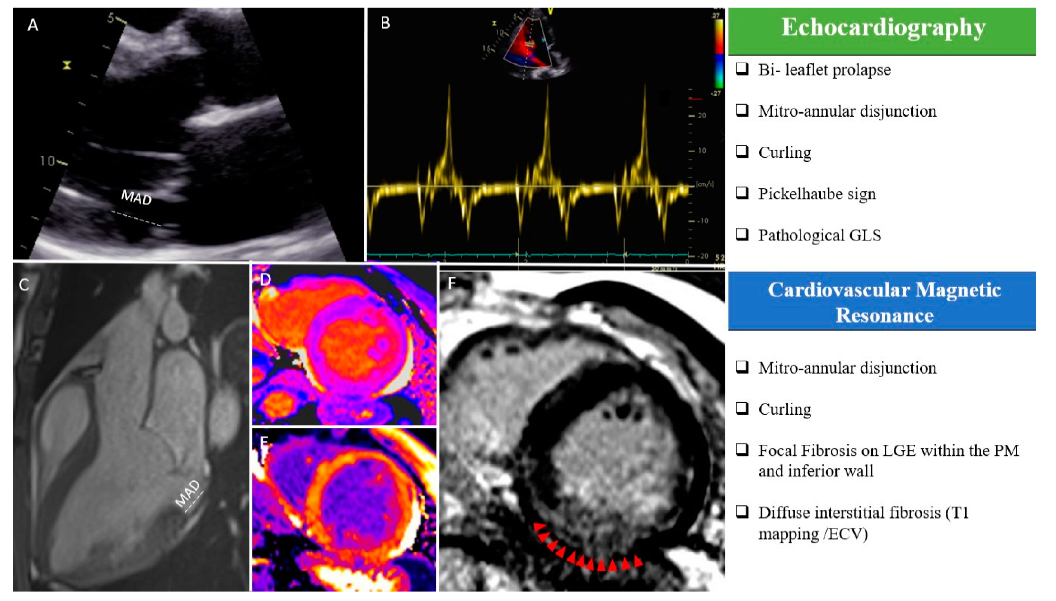
- Bileaflet Prolapse: Myxomatous degeneration involving both leaflets with significant redundancy.
- Mitral Annular Disjunction (MAD): A pathological systolic separation between the posterior mitral valve leaflet hinge point and the basal ventricular myocardium. MAD results in paradoxical systolic expansion of the annulus, exacerbating mechanical stretch. MAD is the anomalous attachment of the posterior leaflet, directly on atrial wall.
- Pickelhaube Sign: On Tissue Doppler Imaging (TDI), a high-velocity systolic signal (S’ ≥ 16 cm/s) at the lateral mitral annulus, reflecting the aggressive traction of the prolapsing leaflets on the annulus.
3. Cardiac Magnetic Resonance (CMR)
CMR is critical for tissue characterization and is highly recommended for AMVP risk stratification, especially in patients with complex arrhythmias or severe MAD.
- Late Gadolinium Enhancement (LGE): The hallmark of the malignant phenotype. Focal LGE is typically found in the papillary muscles or the basal inferolateral mid-myocardium, confirming the presence of the fibrotic arrhythmogenic substrate.
Clinical Management and ICD Indications
Management shifts from simple surveillance to active intervention when the malignant phenotype is suspected.
- Rhythm Monitoring: Extended Holter monitoring (e.g., 7- to 14-day) or implantable loop recorders (ILRs) are strongly indicated for MVP patients with unexplained syncope or significant palpitations to capture silent non-sustained VT (NSVT).
- Medical Therapy: Beta-blockers or non-dihydropyridine calcium channel blockers like verapamil are first-line for suppressing frequent, symptomatic PVCs. Flecainide (if no severe structural heart disease) or Amiodarone may be considered in refractory cases.
- Catheter Ablation: Targeted ablation of the PM or Purkinje network triggers can be highly effective in reducing PVC burden and terminating recurrent VF, though it does not eliminate the need for an ICD if the risk of SCA remains high.
- ICD Implantation:
- Secondary Prevention: Class I indication for MVP patients who have survived SCA or sustained hemodynamically untolerated VT.
- Primary Prevention: Considered (Class IIa/IIb) in patients with AMVP, unexplained syncope, and high-risk features on CMR (e.g., presence of LGE, severe MAD) coupled with complex NSVT on Holter, particularly when EP study is positive or alternative high-risk criteria are met like LVEF <35% and symptomatic HF despite ≥3 months of OMT.
References
Cameron, J. N., Kadhim, K. I., Kamsani, S. H. B., Han, H.-C., Farouque, O., Sanders, P., & Lim, H. S. (2024). Arrhythmogenic Mitral Valve Prolapse: Can We Risk Stratify and Prevent Sudden Cardiac Death? Arrhythmia & Electrophysiology Review, 13. https://doi.org/10.15420/aer.2023.26
Deng, Y., Liu, J., Wu, S., Li, X., Yu, H., Tang, L., Xie, M., & Zhang, C. (2023). Arrhythmic Mitral Valve Prolapse: A Comprehensive Review. Diagnostics, 13(18), 2868. https://doi.org/10.3390/diagnostics13182868
Pavon, A. G., Monney, P., & Schwitter, J. (2021). Mitral Valve Prolapse, Arrhythmias, and Sudden Cardiac Death: The Role of Multimodality Imaging to Detect High-Risk Features. Diagnostics, 11(4), 683. https://doi.org/10.3390/diagnostics11040683
Wu, S., & Siegel, R. J. (2022). Mitral annular disjunction: A case series and review of the literature. Frontiers in Cardiovascular Medicine, 9. https://doi.org/10.3389/fcvm.2022.976066
Sabbag, A., Essayagh, B., Barrera, J. D. R., Basso, C., Berni, A., Cosyns, B., Deharo, J.-C., Deneke, T., Di Biase, L., Enriquez-Sarano, M., Donal, E., Imai, K., Lim, H. S., Marsan, N. A., Turagam, M. K., Peichl, P., Po, S. S., Haugaa, K. H., Shah, D., de Riva Silva, M., Bertrand, P., Saba, M., Dweck, M., Townsend, S. N., & Ngarmukos, T. (2022). EHRA expert consensus statement on arrhythmic mitral valve prolapse and mitral annular disjunction complex in collaboration with the ESC Council on valvular heart disease and the European Association of Cardiovascular Imaging endorsed by the Heart Rhythm Society, by the Asia Pacific Heart Rhythm Society, and by the Latin American Heart Rhythm Society. Europace, 24(12), 1981–2003. https://doi.org/10.1093/europace/euac125
Related Posts


About The Author

Johnson Francis
Former Professor of Cardiology, Calicut Govt. Medical Kozhikode, Kerala, India. Editor-in-Chief, BMH Medical Journal