Pacemaker lead on echocardiogram
Pacemaker lead on echocardiogram
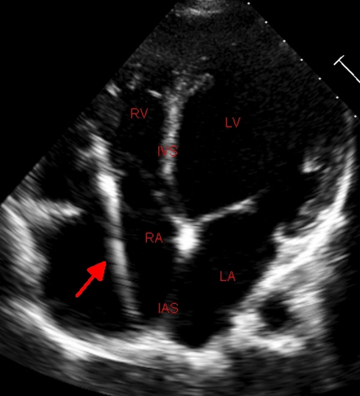
Permanent pacemaker lead is seen extending from the right atrium to the right ventricle with the tricuspid valve in open position. This is an echocardiogram from the apical four chamber view.
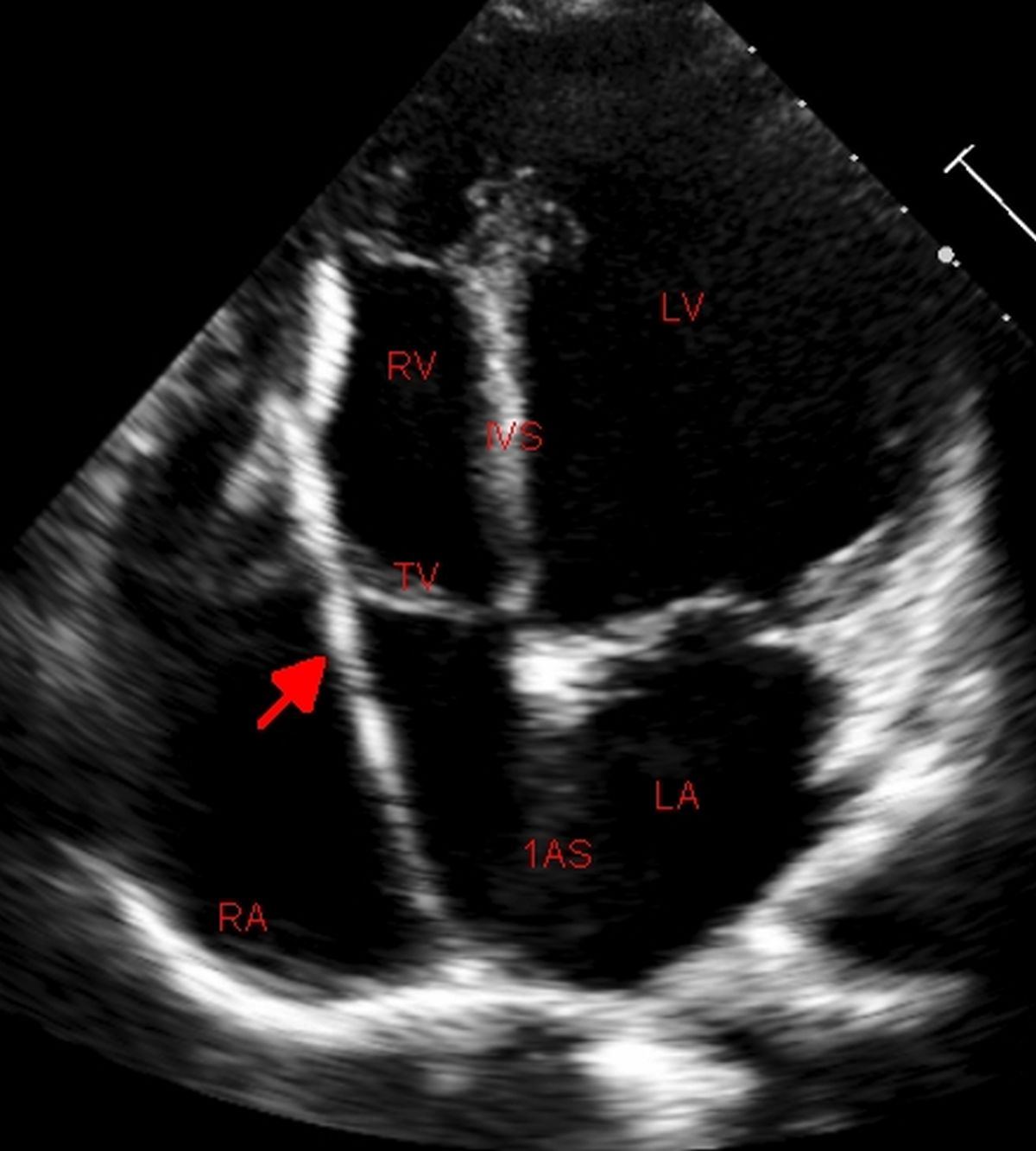
The leaflet marked is the septal leaflet of the tricuspid valve while the leaflet on the other side of the pacemaker lead is anterior tricuspid leaflet. The pacemaker lead is emerging from the upper part of the right atrium from the superior vena cava and it traverses across the closed tricuspid valve into the right ventricle to be inserted in between the coarse trabeculae of the right ventricle.
Echocardiography is useful in detection of pacemaker related complications. In the acute stage it can detect pericardial effusion due to lead perforation. In later stages it can detect vegetations when there is infective endocarditis. Tricuspid regurgitation due to the lead across the tricuspid valve and increase in severity in the long run can be assessed by echocardiography. Left ventricular dyssynchrony and dysfunction secondary to long term right ventricular pacing is also eminently quantified by echocardiography. Thrombus attached to the pacemaker leads can also be detected [1].
Reference
- Almomani A, Siddiqui K, Ahmad M. Echocardiography in patients with complications related to pacemakers and cardiac defibrillators. Echocardiography. 2014 Mar;31(3):388-99.
Related Posts


About The Author
Johnson Francis
Former Professor of Cardiology, Calicut Govt. Medical Kozhikode, Kerala, India. Editor-in-Chief, BMH Medical Journal