Artifacts resembling ventricular tachycardia and fibrillation Artifacts in an ECG can mimic various pathological conditions. In V1, multiple small artefacts almost totally obscure the small QRS complexes so
Mitral regurgitation in PLAX and Apical 4C views Parasternal long axis view showing mitral regurgitation jet into the left atrium. MR is seen as a bluish tongue shaped
Proximal left coronary artery on echocardiogram Proximal portion of major coronary arteries can often be seen on echocardiography, while the distal regions can seldom be imaged. Coronary arteries
Anti-Streptococcal Vaccine: Mutations can occur in weeks, making the vaccine ineffective. Hence an effective anti streptococcal vaccine for prevention of rheumatic fever it difficult to develop.
Severe aortic regurgitation – Echo Severe aortic regurgitation: Echocardiogram in parasternal long axis (PLAX) view shows dilated left ventricle (LV), left atrium (LA), aorta (Ao) and a small
LIMA to LAD coronary bypass graft (CABG) LIMA to LAD coronary bypass graft: LIMA (left internal mammary artery) is to date the most durable conduit for coronary artery
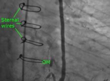
SVG (saphenous vein graft) to obtuse marginal (OM) branch Coronary angiogram in a post coronary artery bypass (CABG) person. Sternal wires indicate the post CABG status. Catheter tip
Bridging collaterals on coronary angiogram Bridging collaterals are a feature of chronic total coronary occlusions. They bridge between the proximal and distal parts of a coronary artery which