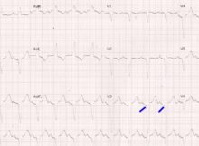
Dock’s murmur in LAD stenosis Dock’s murmur is an early diastolic murmur heard in coronary artery stenosis. Initial documentation was in a man with heart failure due to
Kokeshi phenomenon: stuck rotablator burr Kokeshi phenomenon is a stuck rotablator burr in a heavily calcified coronary artery during attempted rotablation. Though it is a rare complication, it
Reversed mismatch on PET (Positron Emission Tomography) scan is seen in: Left bundle branch block Right ventricular pacing Non ischemic cardiomyopathy All of the above Correct answer: 4)
Tracer used to assess cardiac sympathetic denervation: a) 11C-meta-hydroxyephedrine b) Technetium (99mTc) sestamibi c) Fludeoxyglucose (18F) d) None of the above Correct answer: a) 11C-meta-hydroxyephedrine Myocardial sympathetic denervation assessed by 11C-meta-hydroxyephedrine has been shown
AV block in the presence of features of dilated cardiomyopathy (DCM), may indicate: a) Sarcoidosis b) Lyme disease c) Myotonic dystrophy d) All of the above e) None
Pick the wrong statement: a) Light chain cardiac amyloidosis has a better survival than transthyretin cardiac amyloid amyloidosis b) Light chain cardiac amyloidosis responds to chemotherapy c) Transthyretin
Recommended test for monitoring the effect of bivalirudin during cardiopulmonary bypass: a) Activated clotting time (ACT) b) Ecarin clotting time (ECT) c) Prothrombin time (PT) d) Activated partial
Alternative anticoagulation for a patient with HIT (Heparin induced thrombocytopenia) should not include: a) Low molecular weight heparin b) Warfarin c) Argatroban d) a and b Correct answer: d)