Mechanism/s by which magnesium suppresses torsades de pointes: a) Blockage of L-Type calcium channels b) Suppression of early afterdepolarization c) Reversal of intraventricular dispersion of repolarization d) All
Pseudo atrial fibrillation is seen in:
a) Mitral stenosis
b) Chronic obstructive pulmonary disease
c) Post cardiac transplant state
d) None of the above
Rhythm strip for diagnosis: a) Atrial fibrillation b) Sinus arrhythmia c) Multifocal atrial tachycardia d) None of the above Correct answer: c) Multifocal atrial tachycardia Multifocal atrial tachycardia is
Splintered QRS is a term used in the description of ECG in: a) Ebstein’s anomaly b) Acute myocardial infarction c) Ventricular septal defect d) Pericardial effusion Correct answer: a) Ebstein’s
Factor/s enhancing the risk of digoxin toxicity: a) Co-administration of verapamil b) Hypokalemia c) Hypomagnesemia d) All of the above Correct answer: d) All of the above Hypokalemia which
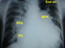
This chest X-ray is suggestive of: a) Primary pulmonary hypertension b) Atrial septal defect with pulmonary hypertension c) Ventricular septal defect with pulmonary hypertension d) Idiopathic dilatation of
Giant wide T inversion may be seen in all except: a) After a cardiac arrest b) Subarachnoid hemorrhage c) Hyperkalemia d) Takotsubo cardiomyopathy Correct answer: c) Hyperkalemia Giant T wave
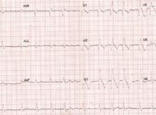
This ECG shows: (Click on the image for an enlarged view) a) Anterior wall infarction b) Anterior wall infarction with right bundle branch block c) Anterior wall infarction