Multiple echo views in EMF Parasternal long axis (PLAX) view shows dilated right ventricular outflow tract. This pattern alone will not give a suspicion of endomyocardial fibrosis as
Parasternal long axis view in normal echocardiogram Parasternal long axis view in normal echocardiogram: RV FW: Right ventricular free wall; RV: Right ventricle; IVS: Interventricular septum; Ao: Aorta;
Mitral regurgitation in PLAX and Apical 4C views Parasternal long axis view showing mitral regurgitation jet into the left atrium. MR is seen as a bluish tongue shaped
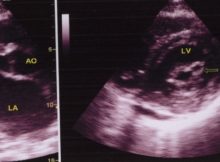
Echocardiogram in Mitral Stenosis Left panel shows the parasternal long axis view (PLAX). AO: Aorta; MVO: Mitral valve opening; LA: left atrium. Doming of the anterior mitral leaflet