Parasternal long axis view in Tetralogy of Fallot – Echocardiogram video
Parasternal long axis view in Tetralogy of Fallot – Echocardiogram video
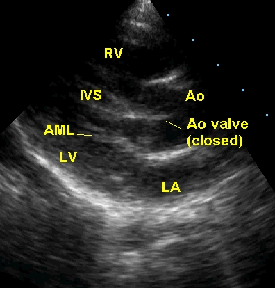
Parasternal long axis (PLAX) view in Tetralogy of Fallot, diastolic frame showing the aortic valve in closed position and mitral valve in open position. The aortic valve appears to impinge on the ventricular septum, but the ventricular septal defect (VSD) with aortic over-ride and connection between right ventricle (RV) and aorta (Ao) is evident just above the septum. LA: left atrium; LV: left ventricle; IVS: interventricular septum; AML: anterior mitral leaflet.
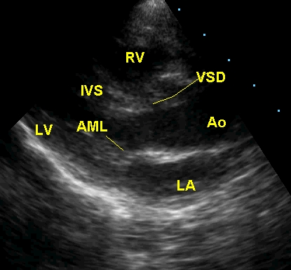
In the systolic frame the VSD with aortic over-ride is quite evident. The mitral valve is in the closed position. This appearance in parasternal long axis view on echocardiography is not specific for Tetralogy of Fallot. The same appearance in this view can occur in pulmonary atresia with ventricular septal defect as well as in truncus arteriosus. Only other views will help us to differentiate between the three conditions.
About The Author
Johnson Francis
Former Professor of Cardiology, Calicut Govt. Medical Kozhikode, Kerala, India. Editor-in-Chief, BMH Medical Journal