Prosthetic heart valves on CXR
Prosthetic heart valves on CXR
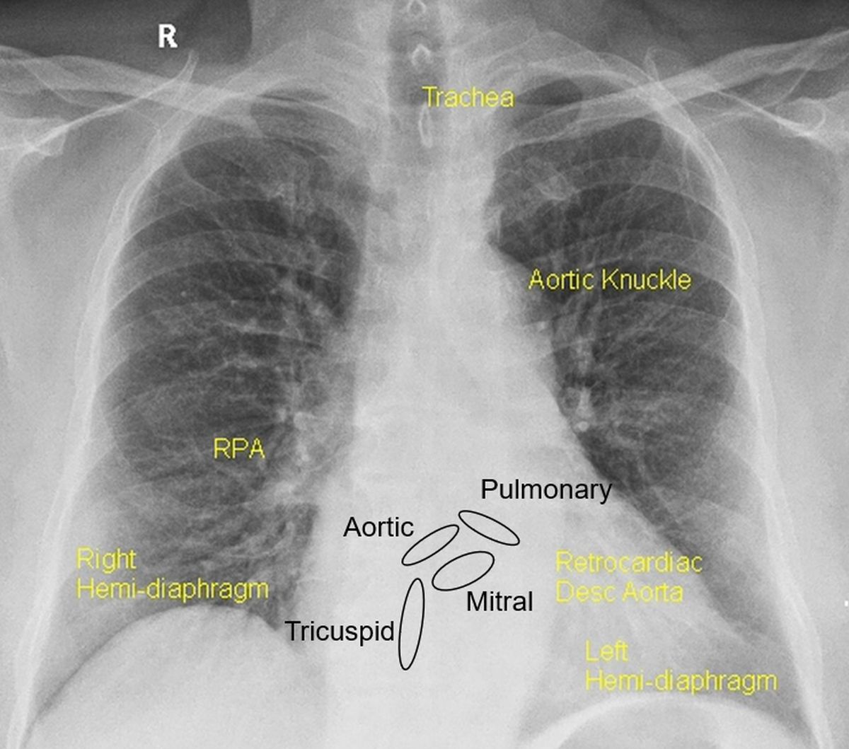
Approximate positions of the valves as seen on the CXR are marked in the picture below. A line connecting the pulmonary bay to the right cardiophrenic angle is used to get the positions of aortic and mitral valves. Mitral valve is below this line while aortic valve is above it. Tricuspid valve is a midline structure. But the actual positions may vary from case to case, especially with differential enlargement of the cardiac chambers. Most commonly replaced valves are the mitral and aortic, tricuspid next and pulmonary the least. Annuloplasty rings used for valve repair may also be seen on CXR.
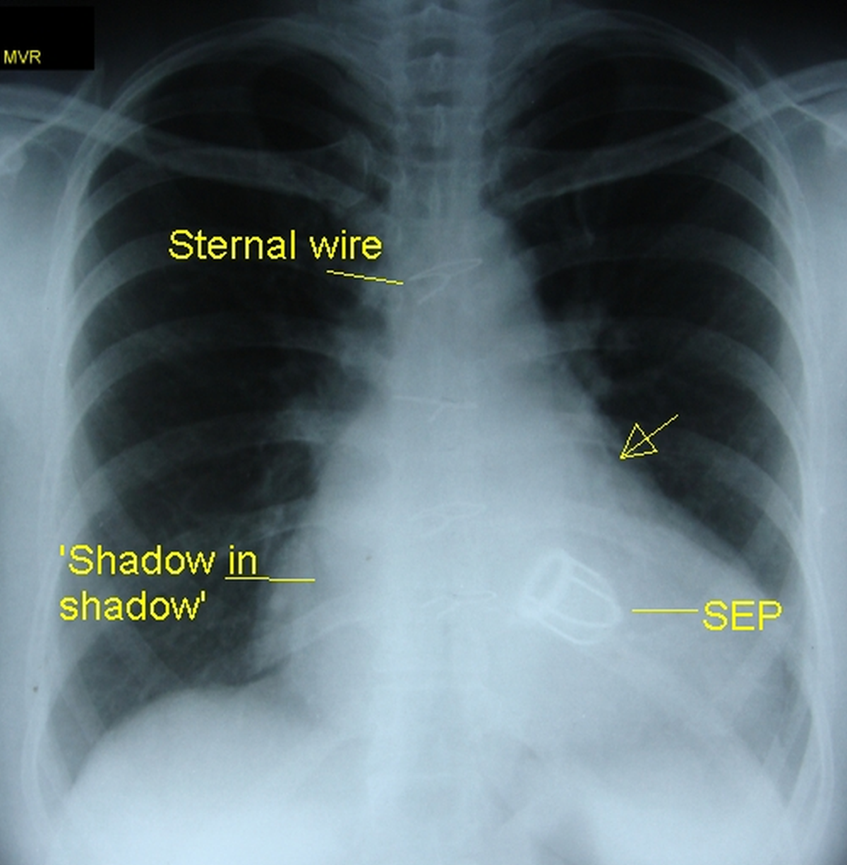
Ball and cage prosthesis: The older valves were of ball and cage design. The prototype was Starr-Edwards valve which was the workhorse with valves functioning well in a person over 5 decades. But due to other problems the valve has been discontinued. The design consists of cage with a sewing ring and struts. Mitral valve had four struts while the aortic had three struts. A ball shaped poppet moved back and forth during cardiac cycle to open and close the valve. Picture below shows a CXR with Starr Edwards prosthetic mitral valve. The valve ring and four struts are visible, but the silastic poppet being non radio opaque, is not visible.
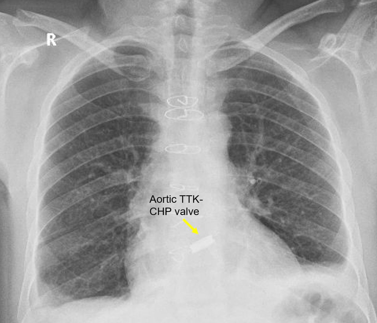
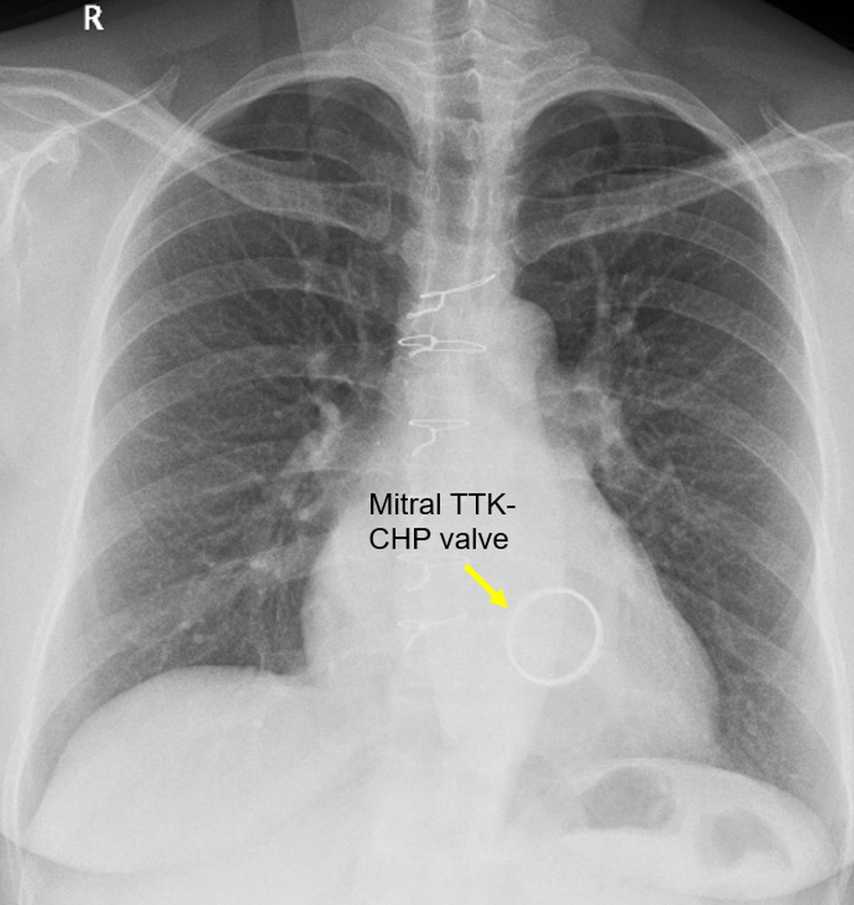
Tilting disc prosthesis: The initial Bjork Shiley valve which has been discontinued was a tilting disc valve. Currently there are single leaflet and bileaflet tilting disc valves. TTK-CHP valve is tilting disc valve with ultra-high-molecular-weight polyethylene disc which is not radiopaque. The valve ring is visible.
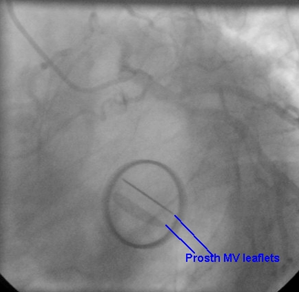
Though prosthetic valves are seen well on CXR, they can be evaluated better by fluoroscopy. In fluoroscopy, valve leaflets which are radio opaque can be seen opening and closing at the corresponding time of the cardiac cycle depending on the position of the valve. A stuck leaflet can be easily identified. Fluoroscopic view of a bileaflet mitral valve is shown below with leaflets in open position.
St. Jude Medical and CarboMedics bileaflet valves have slightly different features, but both have similar clinical performances. Medtronic Hall is a single leaflet valve. On-X is a newer bileaflet valve which has been approved for lesser warfarin requirement in aortic position.
About The Author
Johnson Francis
Former Professor of Cardiology, Calicut Govt. Medical Kozhikode, Kerala, India. Editor-in-Chief, BMH Medical Journal