Suprasternal view in echocardiography
Suprasternal view in echocardiography
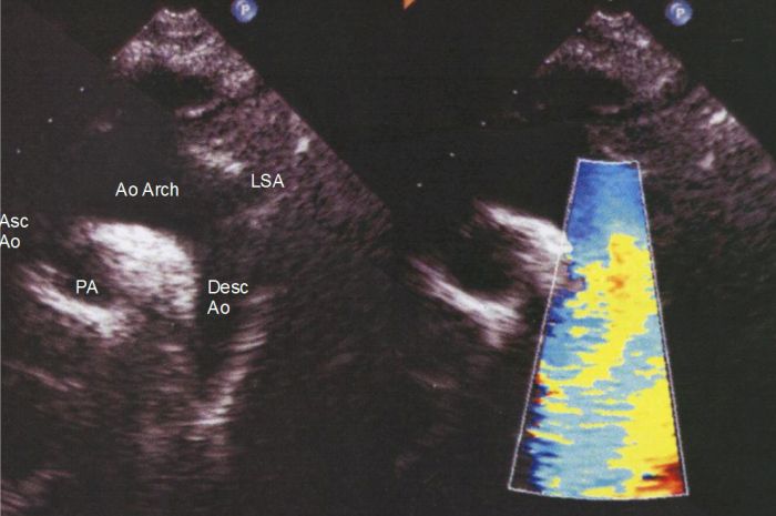
The suprasternal view in echocardiography is obtained by placing the transducer in the suprasternal notch and directing the echo beam downwards towards the heart.
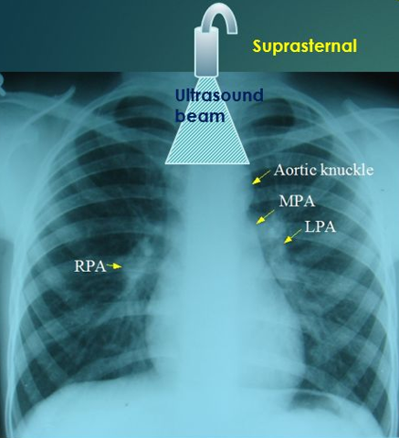
In the adult it can image the arch and portions of the ascending and descending aorta. It can also image the pulmonary artery and the patent ductus arteriosus. In infants, cardiac structures will also be visible in this view. Aorta is always superior in this view and this fact is useful in identifying the relation of the aorta to the pulmonary artery in complex congenital heart disease. The origins of the major arch vessels are also visible in this view. Most often this view is used in congenital heart disease to look for coarctation of aorta. Descending aortic narrowing (usually with a shelf like structure extending from the lateral wall towards the region of the ductus arteriosus) and Doppler gradient are visualized in coarctation of aorta in this view. The ascending aorta can also be imaged by tilting the transducer towards the right. Measurement of aortic gradient in aortic stenosis can be achieved in this view while imaging the ascending aorta, especially with continuous wave (CW) Doppler. Sometimes a pencil probe with CW only imaging helps to pick the gradient of aortic stenosis better in this view.

The right half of the image shows the color flow mapping image in the descending aorta. It shows a predominant blue color due to the flow away from the transducer in the descending aorta. Sometimes the vertical vein can be imaged beyond the descending aorta to the left in hemi anomalous or total anomalous pulmonary venous drainage. The color flow mapping will show red colour in the vertical vein as the flow is upwards to the left brachiocephalic vein. The colour flow will be blue indicating downward flow in the same location if a left superior vena cava is present. The ascending aorta will show a red flow and the right superior vena caval flow may be imaged, especially in children as a blue color.
Related Posts

About The Author
Johnson Francis
Former Professor of Cardiology, Calicut Govt. Medical Kozhikode, Kerala, India. Editor-in-Chief, BMH Medical Journal