Rheumatic heart disease This lecture will discuss about rheumatic fever, latest diagnostic criteria (Revised by American Heart Association in 2015), and an overview of rheumatic heart disease. Echocardiogram
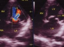
Doppler jet in mitral stenosis with regurgitation Doppler jet in mitral stenosis with regurgitation: Mitral Doppler interrogation is usually done from the apical four chamber view. Good colour
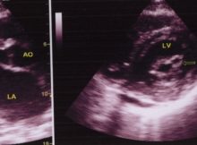
PLAX view in MS Echocardiogram in parasternal long axis view in mitral stenosis. The anterior mitral leaflet is seen to be domed, with a hockey stick appearance. The
Color Doppler Echocardiogram in Mitral Stenosis showing thickened and domed mitral leaflets, turbulent colour jet across the mitral valve in the right panel and Doppler tracing with E
Mechanism of loud first heart sound in mitral stenosis In severe mitral stenosis, due to elevated left atrial pressure, the closure of the mitral valve is delayed and
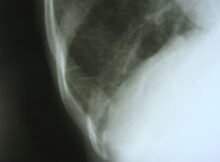
Straightening of left border in mitral stenosis, on X-ray chest PA view The uppermost portion on the left cardiac border is the aortic knuckle. The next slight bulge
Echocardiogram in Mitral Stenosis Left panel shows the parasternal long axis view (PLAX). AO: Aorta; MVO: Mitral valve opening; LA: left atrium. Doming of the anterior mitral leaflet
Wilkins echocardiographic score for mitral stenosis Wilkins echocardiographic score for mitral stenosis is useful for deciding whether the valve is suitable for balloon mitral valvotomy (BMV), also known