Transesophageal echocardiogram in atrial septal defect – Echocardiogram video
Transesophageal echocardiogram in atrial septal defect – Echocardiogram video
Transoesophageal echocardiogram (TEE) is useful in the evaluation of atrial septal defect (ASD) to assess the finer details while deciding on device closure. It is also useful in delineating ASDs which are not visible by transthoracic echocardiography (TTE) either due to poor echo window or due to odd location of the ASD as in sinus venosus ASD. TEE is often used in this context while evaluating pulmonary hypertension of obscure etiology in an adult. TEE probe being very near the heart without any intervening lung tissue, can give excellent images. Moreover, the short distance permits the use of higher frequency transducers with better image resolution. Usually higher frequency transducers cannot be used for TTE because of poor depth of ultrasound penetration at higher frequencies in an adult.
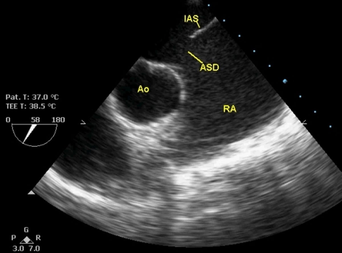
TEE image in short axis view showing the aorta (Ao), part of the interatrial septum (IAS) and the ASD. It can be seen that there is hardly any aortic rim (bald aortic rim). Part of the left atrium is seen above the IAS at the top (not marked in the figure). Below the IAS the large right atrium is visible. The TEE probe has a temperature sensor which senses both the subjects temperature as well as the probe temperature. Automatic cooling is initiated when the probe temperature goes above the cut off limit. A visible alert is also displayed when the probe heats up, requesting the operator to reduce the power output of the ultrasound beam. A trade off between visibility of images and the heating up of the probe is required in some cases. Since it is difficult to repeat TEE studies frequently, either continuous or frequent recording of TEE videos during the entire study will permit reassessment by the same operator as well as an independent operator. TEE studies are invariably done in a fasting state and under topical anaesthesia of the oropharynx to reduce gag reflex. A mouth gag is needed to prevent biting and damage to the probe. ECG monitoring is needed to time the events in the cardiac cycle as well for the rhythm in sick individuals. Pulse oximetry and facility for suction are also ideal. Pat. T: temperature of the subject; TEE T: TEE probe temperature. The dial shows the plane of imaging from 0-180 degrees (58 degrees in this image).

Dual ASDs with a small intervening segment of atrial septum seen on TEE. The right atrium appears dilated. The total size of both ASDs taken together is quite large and not suitable for device closure.
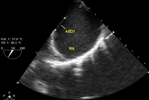
First of the two ASDs being measured by calipers. The next image shows the measurements of both ASDs.
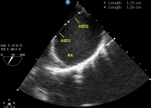
This image displays the measurements of both ASDs. One measures 17.5 mm and another measures 15.6 mm. The total will be 33.1 mm. The rims at both ends also appear deficient so that device closure may not be feasible in this case. Surgical closure will be ideal, provided that there is no features of irreversible pulmonary hypertension.
Related Posts


About The Author
Johnson Francis
Former Professor of Cardiology, Calicut Govt. Medical Kozhikode, Kerala, India. Editor-in-Chief, BMH Medical Journal