Vegetation on tricuspid valve – echocardiographic image
Vegetation on tricuspid valve – echocardiographic image
Vegetation on tricuspid valve – echocardiographic image:
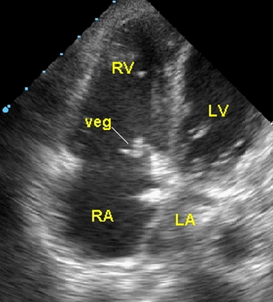
Vegetation on tricuspid valve seen on echocardiography from a modified apical four chamber view. LV: left ventricle; LA: left atrium; RV: right ventricle; RA: right atrium. In live echo imaging, the vegetation will show a motion pattern independent of the movement of the tricuspid leaflet. Tricuspid valve endocarditis with vegetation is seen in intravenous drug users or occasionally in those with a central venous catheter in situ for a long period. A similar situation is a transjugular hemodialysis catheter. When a catheter is associated with vegetation it is usually removed and the tip sent for culture. Blood cultures as per standard protocol are also taken along with it. Tricuspid valve vegetations can embolize into the pulmonary circulation leading to septic pulmonary embolism. Xin Yu Song, Shan Li, Jian Cao, Kai Xu, Hui Huang and Zuo Jun Xu noted in their retrospective study of 20 cases of cardiac septic pulmonary embolism that there were right sided vegetations in 15 cases [1].
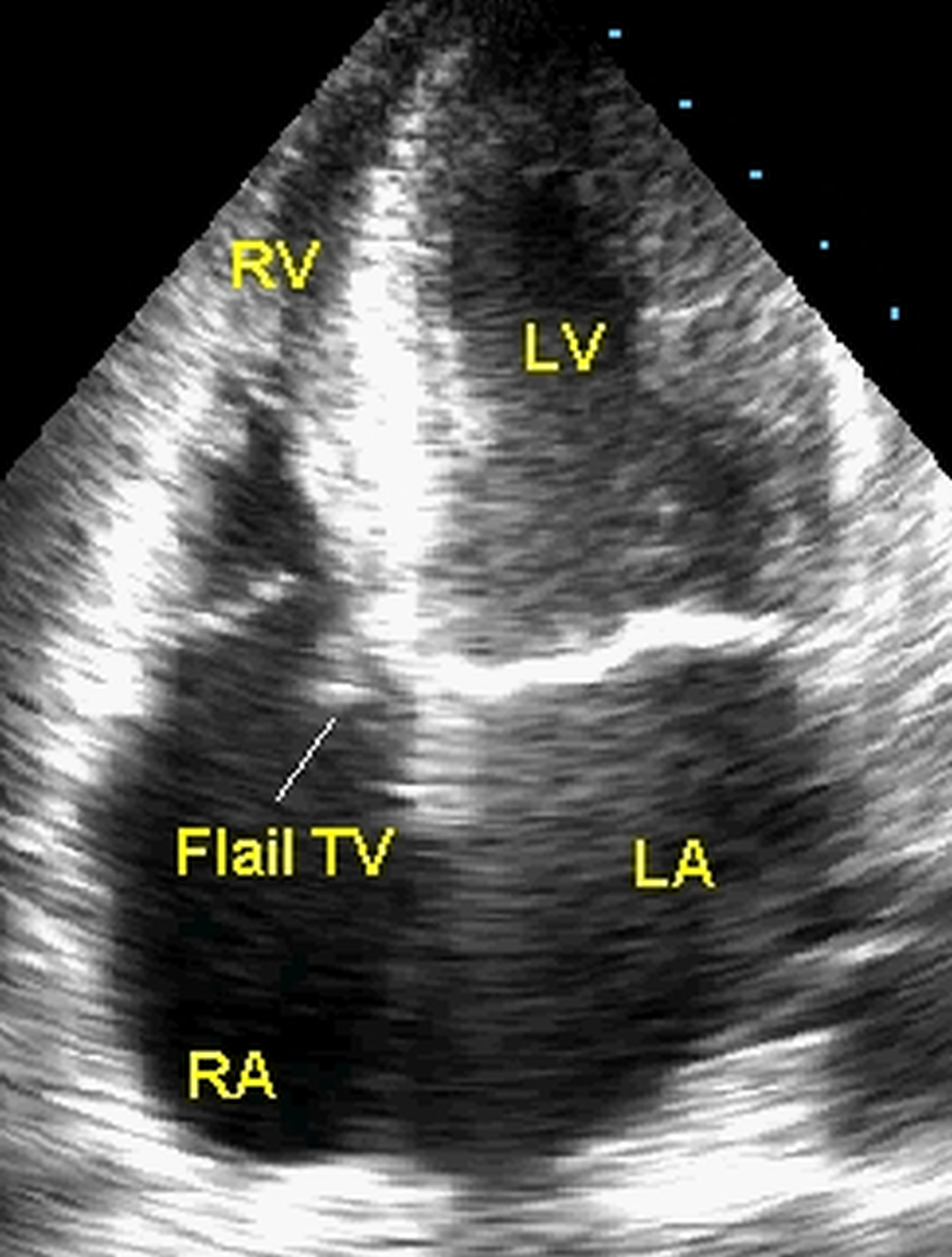
Repeat echo at a later date after antibiotic treatment showing regression of vegetation and a flail septal tricuspid leaflet pointing towards the right atrium. Flail leaflet is a result of rupture of the chordae tendineae and causes tricuspid regurgitation as demonstrated below.
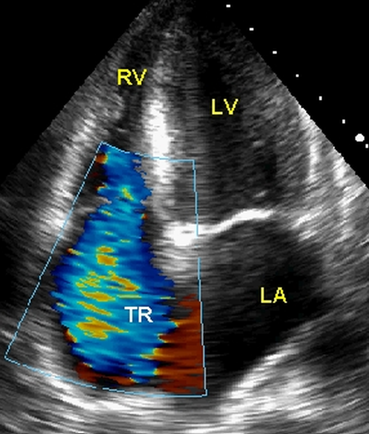
Severe tricuspid regurgitation seen as a bluish mosaic (multi-color) jet from right ventricle to right atrium in systole. The mitral valve can be seen in the closed position between left ventricle and left atrium indicating that it is a systolic frame.
Reference
- Xin Yu Song, Shan Li, Jian Cao, Kai Xu, Hui Huang, Zuo Jun Xu. Cardiac septic pulmonary embolism: A retrospective analysis of 20 cases in a Chinese population. Medicine (Baltimore). 2016 Jun;95(25):e3846.
Related Posts

About The Author
Johnson Francis
Former Professor of Cardiology, Calicut Govt. Medical Kozhikode, Kerala, India. Editor-in-Chief, BMH Medical Journal