What is ECG imaging (ECGi)?
What is ECG imaging (ECGi)?
ECG imaging (ECGi) is a non-invasive three dimensional electrophysiology mapping using a 252 electrode system applied as a vest over the chest. CardioInsight™ uses a single use, disposable, multi-electrode mapping vest to acquire electrophysiological data from the body surface. It combines the surface ECG signals with computed tomography (CT) scan data to produce and display simultaneous 3-D cardiac maps from all four cardiac chambers [1].
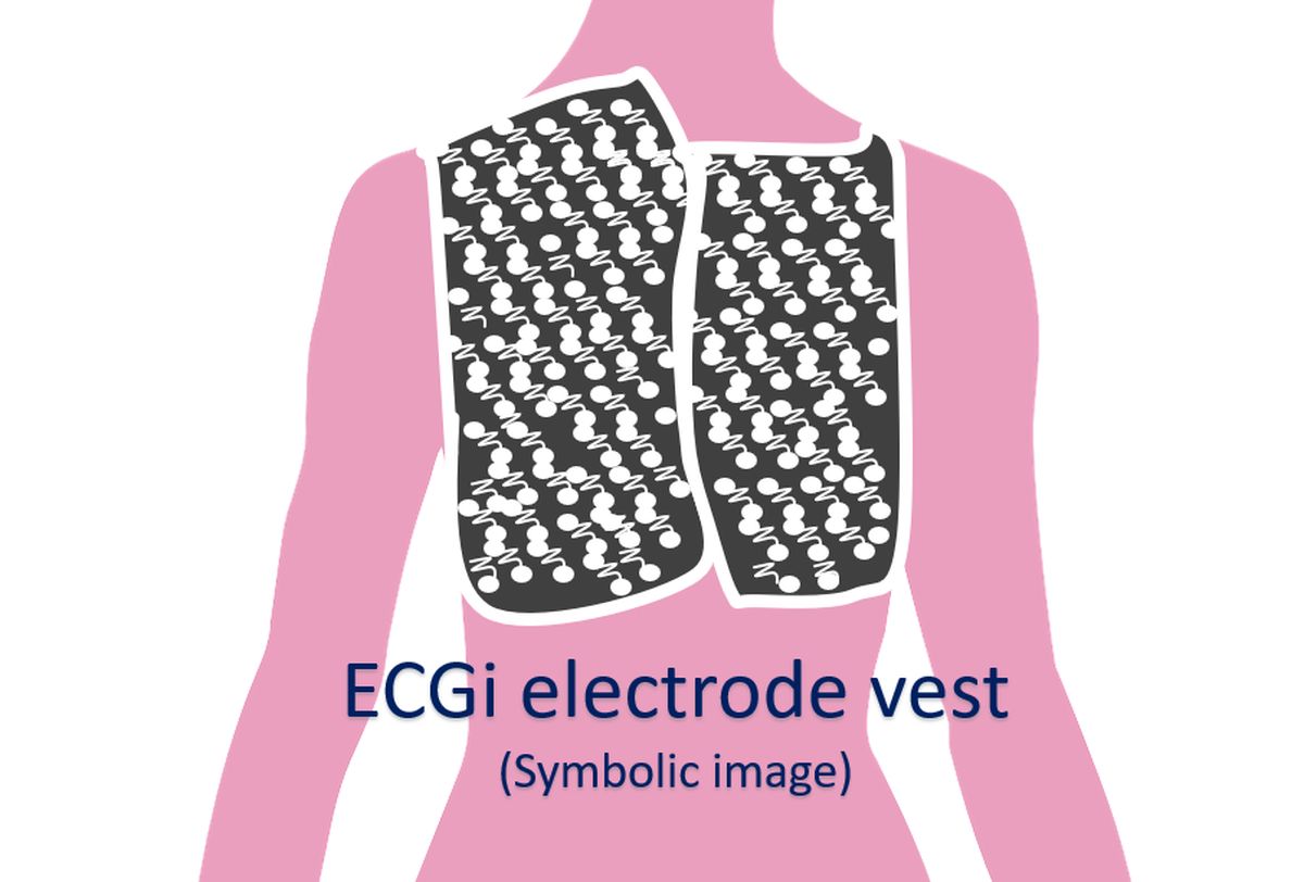
The vest has three multi-electrode panels for front left, front right and back. Connectors from the electrode vest is connected to the CardioInsight workstation. The chest is shaved and prepared with non-alcoholic, hypoallergenic solution. The front left panel is applied first followed by the front right and back panels. Panels are secured with medical tape. CT scan is done with vest on. Signals from the 252 electrodes are obtained and integrated with the CT data. Alternatively, the ECG data can be acquired first and CT images obtained shortly after. The workstation provides various types of maps including phase map, composite map, potential map, activation map, propagation map, voltage map, slew rate map as well as virtual electrograms.
ECGi constructs electrograms over the entire epicardial surface of the heart with high resolution. Local activation and recovery times can be determined from the electrograms. Activation-recovery intervals (a measure of local action potential duration) can be computed from ECGi data. Activation-isochrone maps and repolarization pattern maps of the heart surface can be obtained noninvasively by ECGi [2].
ECGi uses a standardized workflow and it has been suggested that signals should be checked manually to avoid automatic processing errors. Presence of local activation covering the arrhythmia cycle length confirms re-entry. QS pattern associated with centrifugal activation demonstrates focal breakthrough. ECGi helps us to understand the mechanism of cardiac arrhythmias which could guide catheter ablation [3].
ECGi has been used to exclusively guide ablation of premature ventricular complexes [4]. The utility of ECGi to map hemodynamically stable and unstable ventricular arrhythmias has been studied. In that study simultaneous electroanatomic mapping (CARTO, Biosense-Webster) and ECGi (CardioInsight, Medtronic) were done in 18 patients during catheter ablation of ventricular tachycardia (VT). A total of 29 VTs were available for comparison. ECGi mapped VT sites of origin correctly with higher accuracy than a validated 12-lead ECG algorithm (83.3% vs 38.9%). It was shown that CardioInsight localizes VT circuits with sufficient accuracy to provide a region of interest for targeting mapping for ablation. But the resolution was not enough to guide discrete radiofrequency lesion delivery by catheter ablation. The authors suggested that the resolution may be sufficient to guide segmental ablation with stereotactic radiotherapy [5].
ECGi has been used to identify rotational activity non-invasively that can be targeted during catheter ablation of atrial fibrillation. This has been used to predict procedural outcome in persistent and long-standing persistent atrial fibrillation. Acute termination using rotational ablation guided by ECGi phase mapping could be achieved in a high percentage of both index and re-do cases [6].
Noninvasive electroanatomic mapping (NIEAM, CardioInsight, Medtronic) can demonstrate patterns of depolarization that are useful in identifying the chamber of origin in outflow tract ventricular arrhythmias. But arrhythmia breakout and signal directionality had poor diagnostic value in predicting site of origin in outflow tract ventricular arrhythmias and underperformed compared to ECG interpretation (59.1% and 80.5%). After excluding instantaneous unipolar electrograms with poor characteristics, the instantaneous unipolar electrograms with most negative amplitude at the chamber of origin was predictive of the true site of origin with 96.4% sensitivity and specificity [7].
References
- Medtronic CardioInsight™ Mapping Vest. Available at: https://europe.medtronic.com/xd-en/healthcare-professionals/products/cardiac-rhythm/cardiac-mapping/cardioinsight-mapping-vest.html. Accessed on 2 April, 2022.
- Rudy Y. Noninvasive Mapping of Repolarization With Electrocardiographic Imaging. J Am Heart Assoc. 2021 May 4;10(9):e021396. doi: 10.1161/JAHA.121.021396. Epub 2021 Apr 21. PMID: 33880937; PMCID: PMC8200737.
- Cheniti G, Puyo S, Martin CA, Frontera A, Vlachos K, Takigawa M, Bourier F, Kitamura T, Lam A, Dumas-Pommier C, Pillois X, Pambrun T, Duchateau J, Klotz N, Denis A, Derval N, Cochet H, Sacher F, Dubois R, Jais P, Hocini M, Haissaguerre M. Noninvasive Mapping and Electrocardiographic Imaging in Atrial and Ventricular Arrhythmias (CardioInsight). Card Electrophysiol Clin. 2019 Sep;11(3):459-471. doi: 10.1016/j.ccep.2019.05.004. PMID: 31400870.
- Erkapic D, Neumann T. Ablation of premature ventricular complexes exclusively guided by three-dimensional noninvasive mapping. Card Electrophysiol Clin. 2015 Mar;7(1):109-15. doi: 10.1016/j.ccep.2014.11.010. Epub 2014 Dec 12. PMID: 25784027.
- Graham AJ, Orini M, Zacur E, Dhillon G, Daw H, Srinivasan NT, Martin C, Lane J, Mansell JS, Cambridge A, Garcia J, Pugliese F, Segal O, Ahsan S, Lowe M, Finlay M, Earley MJ, Chow A, Sporton S, Dhinoja M, Hunter RJ, Schilling RJ, Lambiase PD. Evaluation of ECG Imaging to Map Hemodynamically Stable and Unstable Ventricular Arrhythmias. Circ Arrhythm Electrophysiol. 2020 Feb;13(2):e007377. doi: 10.1161/CIRCEP.119.007377. Epub 2020 Jan 14. PMID: 31934784.
- Gao X, Lam AG, Bilchick KC, Darby A, Mehta N, Mason PK, Malhotra R, Mangrum JM. The use of non-invasive mapping in persistent AF to predict acute procedural outcome. J Electrocardiol. 2019 Nov-Dec;57S:S21-S26. doi: 10.1016/j.jelectrocard.2019.08.012. Epub 2019 Aug 20. PMID: 31474375.
- Coleman KM, Saleh M, Makker P, Vaishnav AS, Atteya G, Shein J, Bhullar A, Skipitaris NT, Mountantonakis SE. Surface unipolar electrogram characteristics to predict site of origin of outflow tract arrhythmias using noninvasive mapping. J Cardiovasc Electrophysiol. 2021 Feb;32(2):391-399. doi: 10.1111/jce.14857. Epub 2021 Jan 6. PMID: 33368754.
Related Posts

About The Author
Johnson Francis
Former Professor of Cardiology, Calicut Govt. Medical Kozhikode, Kerala, India. Editor-in-Chief, BMH Medical Journal