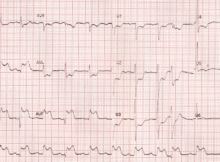
Best treatment option? ECG Quiz What would be the best treatment option for the person with this ECG? ECG shows upsloping ST segment elevation in inferior leads II,
Same day discharge for peripheral arterial procedures in elderly: SENIOR-ER registry Peripheral arterial procedures in elderly: Same day discharge after peripheral arterial procedures have been documented to be safe
Multiple arterial grafts give better long term results A Canadian study [1] published in JAMA Cardiology has shown lower long term mortality, repeat revascularization, myocardial infarction and heart failure in
Long term efficacy of ICD in Brugada Syndrome So far the only proven therapy to prevent sudden cardiac death in Brugada syndrome is the implantation of an ICD,
Hypertrophic cardiomyopathy mutation corrected in embryo In a pathbreaking proof of concept experimental study, MYBPC3 gene mutation causing hypertrophic cardiomyopathy was corrected in human embryos using CRISPR-Cas9 gene
Initiation and spontaneous termination of SVT Initial monitor screen shot shows initiation of SVT and the second one shows spontaneous termination of SVT. SVPC to SVT Monitor screenshot showing initiation
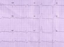
ECG Quiz 14 – Choose the best answer: a) Left bundle branch block b) Right ventricular apical pacing c) AV sequential pacing d) Left bundle branch block with
CRT-D better than CRT-P in NIDCM with left ventricular midwall fibrosis CRT-D better than CRT-P in NIDCM: Francisco Leyva and colleagues compared the outcomes of cardiac resynchronization therapy
Continuous wave Doppler tracing from apical five chamber view Continuous wave Doppler tracing showing aortic stenosis and regurgitation jets. The aortic regurgitation jet has a rapid rise in