ASD on colour Doppler echocardiogram Subcostal view showing a 15 mm ASD on colour Doppler echocardiogram SVC flow: Flow from superior vena cava into the right atrium. ASD
Distal LAD filling from homocollaterals – coronary angiogram Coronary angiogram showing distal LAD filling from homocollaterals Left coronary angiogram showing total occlusion of left anterior descending coronary artery
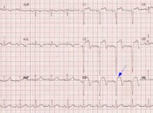
ECG filter settings and detection of pacemaker spikes In modern day digital ECGs, ECG filter settings can beautify the tracings, but vital information can be filtered out, as
Pictorial review of CRT implantation Left subclavian venogram – First step in CRT implantation! CRT implantation: Left subclavian venogram is usually the first step during the implantation of
Atrial pacing ECG ECG showing atrial pacing artefact before each P wave Pacing artefacts (stimulus artefacts or pacing spikes) are seen before each P wave. P waves have