STEMI – Anterior Wall
STEMI – Anterior Wall
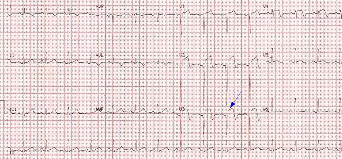
ECG shows ST elevation myocardial infarction of anterior wall. Up sloping elevation of ST segment is seen in leads V1 to V5, with maximum ST elevation in V2, as is characteristic of anterior wall infarction. T waves are inverted in leads V2 to V5 suggesting that it is an evolved myocardial infarction rather than in the very early phase. T wave inversions are seen in leads I and aVL as well. There is no reciprocal ST segment depression in the inferior leads, possibly because it is beyond the acute phase. Similar changes may also be noted in left ventricular aneurysm after a myocardial infarction as the ST segment may remain elevated when there is a dyskinetic segment. QS complexes are seen from V1 to V3. Rounded negative P waves in V1 would suggest associated left ventricular dysfunction causing left atrial overload.
Related Posts


About The Author
Johnson Francis
Former Professor of Cardiology, Calicut Govt. Medical Kozhikode, Kerala, India. Editor-in-Chief, BMH Medical Journal