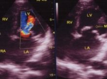
Mitral regurgitation jet by color Doppler echocardiography. Vena contracta is the narrowest portion of the MR jet at or just beyond the regurgitant orifice. It is an accurate
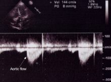
Aortic flow velocity by Doppler echo is routinely measured and is useful to assess aortic gradient in aortic stenosis. It is also useful in calculating valve area by
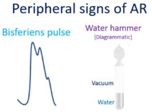
Peripheral signs of aortic regurgitation (AR) are mostly due to the high stroke volume and high pulse pressure. These are features of aortic run off and can occur
Color Doppler Echocardiogram in Mitral Stenosis showing thickened and domed mitral leaflets, turbulent colour jet across the mitral valve in the right panel and Doppler tracing with E
Ebstein’s Anomaly of Tricuspid Valve Parasternal short axis view in Ebstein’s anomaly, showing the dilated right ventricular outflow tract (RVOT). Ao: Aorta. Parasternal long axis view in Ebstein’s
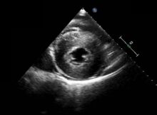
LVH on echocardiogram Parasternal short axis view at the level of the papillary muscles showing severe concentric left ventricular hypertrophy. Serial short axis views have to be obtained
D shaped LV cavity in severe RVH Echo from parasternal short axis view shows a D shaped LV cavity (left ventricular cavity) alongside a grossly hypertrophied right ventricle
M-Mode echocardiogram at Aorta – Left atrium level M-Mode echocardiogram at Aorta – Left atrium level: M-Mode cuts are usually obtained from two dimensional (2-D) views after the proper