Atrial septal defect with right to left shunt should prompt us to look for total anomalous pulmonary venous connection as well as severe pulmonary hypertension.
Transesophageal Echocardiography (TEE) Transesophageal Echocardiography (TEE) is a specialised form of echocardiographic study using an echo probe held at the tip of an endoscope like device. As the
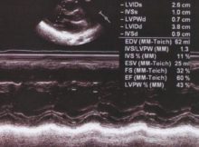
M-Mode echocardiography and Anatomical M-Mode M-Mode echocardiography (Time-motion mode) was one of the earliest tools of the echocardiographer. M-Mode gives an ice-pick view of the heart. The vertical
Left atrial myxoma – echocardiogram with video in parasternal long axis view with systolic and diastolic frames. Mitral regurgitation is also seen on color Doppler.
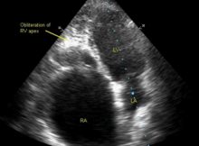
Diagnosis based on the echocardiogram: a) Ebstein’s anomaly of tricuspid valve b) Idiopathic dilatation of right atrium c) Endomyocardial fibrosis d) None of the above Correct Answer
60/60 sign has been described in: a) Acute myocardial infarction b) Acute aortic dissection c) Acute pulmonary embolism d) None of the above Correct Answer
Aorta – left atrium M-mode Mode echocardiogram at aorta-left atrium level Mode tracing at the aorta-left atrium level is often the first M-mode tracing to be taken from the
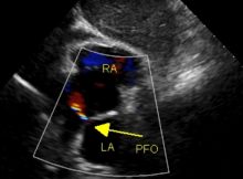
Patent foramen ovale – PFO Patent foramen ovale is a valvular opening in the fossa ovalis region of the interatrial septum. In fetal life, the foramen ovale shunts