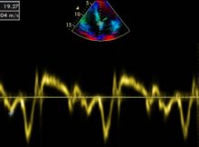
Diastolic dysfunction by tissue Doppler E/E’ measurement by Tissue Doppler Imaging E/E’ measurement is used to assess diastolic function by tissue Doppler. Method may vary in technical details
Recommended view for measuring left ventricular (LV) thickness by echocardiography in hypertrophic cardiomyopathy (HCM):
a) Parasternal long axis view by 2D
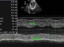
b) Parasternal long axis view by M-Mode
c) Short axis
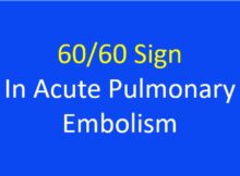
Echocardiography is useful in demonstrating right heart thrombi, features of right ventricular overload and dysfunction as well as in differential diagnosis of acute pulmonary embolism.
60/60 sign in acute pulmonary embolism combines tricuspid regurgitation gradient of less than 60 mm Hg and pulmonary flow acceleration time of less than 60 msec.
IVC plethora is seen in all of the following except:
a) Acute left ventricular failure
b) Chronic right heart failure
c) Cardiac tamponade
d) Constrictive pericarditis
Conventionally, aortic stenosis has been classified into mild (peak gradient up to 50 mm Hg), moderate (peak gradient between 50 - 75 mm Hg) and severe, with